Large Synthetic Moissanite with Silicon Carbide Polytypes
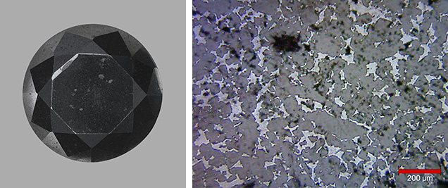
Silicon carbide (SiC) is composed of a carbon atom surrounded by four silicon atoms in a tetrahedral form. Colorless synthetic moissanite is pure silicon carbide; the incorporation of impurities produces colors, including black. Its high hardness (second only to diamond) and thermal conductivity make it a convincing diamond simulant. Both transparent and opaque synthetic moissanites have been submitted to GIA’s laboratory over the past two decades. In this report, we examine a 29.73 ct synthetic moissanite submitted as a black diamond (figure 1, left).
An uneven surface and surface-breaking black inclusions were visible with the unaided eye. Mosaic patterns of light and dark gray regions were observed under a microscope and became distinct in the Raman image (figure 1, right). Small cavities and polish lines in different directions were present on the table surface, and a granular texture was observed in a large cavity on the girdle. Raman spectroscopy identified the dark gray regions as silicon carbide.
Interestingly, each random testing spot showed mixed bands of silicon carbide polytypes (i.e., identical chemical composition but a slightly different crystal structure). The polytype 6H-SiC (6 = the number of stacking sequences, H = hexagonal, and SiC = silicon carbide) was detected by Raman bands at 789.6, 765.0, and 148.0 cm–1. Other polytypes were also distinguished by Raman bands: 3C-SiC (C = cubic) at 970.1, 968.4, 797.8, 795.4, and 788.2, cm–1; 4H-SiC at 785.7 cm–1; and 15R-SiC (R = rhombohedral) at 768.4 cm–1 (figure 2; see also P. Colomban, “SiC, from amorphous to nanosized materials, the example of SiC fibers issued of polymer precursors,” in M. Mukherjee, Ed., Silicon Carbide—Materials, Processing and Applications in Electronic Devices, InTech, 2011, pp. 161–186). In addition to these bands, a sharp Raman band assigned to silicon was detected at 520.2 cm–1 at a few spots. Bands corresponding to 6H-SiC at 238.8 and 148.0 cm–1 could not be detected at all measurement locations. Light gray regions were identified as silicon at 520.0 cm–1 (figure 2, left inset). Black inclusions, both eye-visible and microscopic, were confirmed as graphite. As figure 3 illustrates, the micro-image of a graphite inclusion showed an amorphous graphite vein (broad Raman bands at approximately 1561.0 and 1339.0 cm–1) inside a crystalline graphite matrix (sharp bands at 1580.0 and 1350.0 cm–1)


During SiC growth, rotation along the covalent bond generated alternate stacking layers of Si and C atoms along the c-axis with different structural sequence, which is noticeable in the (1120) plane but not along the basal plane. This rotation can occur at very low energy. Since constant growth energy is difficult to control, two or more silicon carbide polytypes can form simultaneously during the growth process; this phenomenon is called polytypism. Raman spectroscopy has been used to detect these polytypes. Although there are more than 250 known polytypes, SiC can be classified as either beta (β) or alpha (α) type. The beta type crystallizes in cubic lattice symmetry (e.g., 3C-SiC), the alpha type in hexagonal symmetry (e.g., 6H-SiC).
Traditional growth methods for silicon carbide involve sintering and hot pressing, and there are also newer techniques. Reaction-bonded silicon carbide can form when liquid silicon reacts with porous graphite. This silicon carbide contains both pure silicon (as a bonding component), and graphite. Earlier studies have suggested that black synthetic moissanite containing silicon inclusions could be grown by the physical vapor transport (PVT) method (see Winter 2009 GNI, p. 308; Spring 2011 Lab Notes, pp. 54–55). Using PVT, researchers have grown large silicon carbide bulk crystals up to 50 × 25 mm at a rate of 1.2 mm per hour at 10 kPa pressure (Q.-S. Chen et al., “Growth of silicon carbide bulk crystals by physical vapor transport method and modeling efforts in the process optimization,” Journal of Crystal Growth, Vol. 292, 2006, pp. 197–200). Nevertheless, Raman analysis at several testing points confirmed only the 6H-SiC polytype.
This sample was submitted for a Colored Diamond Identification and Origin Report, but one should always pay close attention to surface features, such as the mosaic pattern of light and dark gray regions in reflected light, together with surface-breaking black inclusions not observable in black diamond.


