Gem Characterization

The Formation of Natural Type IIa and IIb Diamonds
Evan M. Smith and Wuyi Wang
GIA, New York
Many of the world’s largest and most valuable gem diamonds exhibit an unusual set of physical characteristics. For example, in addition to their conspicuously low nitrogen concentrations, diamonds such as the 3,106 ct Cullinan (type IIa) and the Hope (type IIb, boron bearing) tend to have very few or no inclusions, and in their rough state they are found as irregular shapes rather than as sharp octahedral crystals. It has long been suspected that type IIa and IIb diamonds form in a different way than most other diamonds.
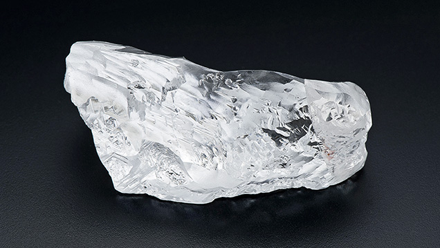
Over the past two years, systematic investigation of both type IIa and IIb diamonds at GIA has revealed that they sometimes contain rare inclusions from unique geological origins. Examination of more than 130 inclusion-bearing samples has established recurring sets of inclusions that clearly show many of these diamonds originate in the sublithospheric mantle, much deeper in the earth than more common diamonds from the cratonic lithosphere. We now recognize that type IIa diamonds, or more specifically, diamonds with characteristics akin to the historic Cullinan diamond (dubbed CLIPPIR diamonds), are distinguished by the occurrence of iron-rich metallic inclusions. Less frequently, CLIPPIR diamonds also contain inclusions of majoritic garnet and former CaSiO3 perovskite that constrain the depth of formation to within 360–750 km. The inclusions suggest that CLIPPIR diamonds belong to a unique paragenesis with an intimate link to metallic iron in the deep mantle (Smith et al., 2016, 2017). Similarly, findings from type IIb diamonds also place them in a “superdeep” sublithospheric mantle setting, with inclusions of former CaSiO3 perovskite and other high-pressure minerals, although the iron-rich metallic inclusions are generally absent (Smith et al., 2018). Altogether, these findings show that high-quality type II gem diamonds are predominantly sourced from the sublithospheric mantle, a surprising result that has refuted the notion that all superdeep diamonds are small and non-gem quality. Valuable information about the composition and behavior of the deep mantle is cryptically recorded in these diamonds. CLIPPIR diamonds (figure 1) confirm that the deep mantle contains metallic iron, while type IIb diamonds suggest that boron and perhaps water can be carried from the earth’s surface down into the lower mantle by plate tectonic processes. In addition to being gemstones of great beauty, diamonds carry tremendous scientific value in their unique ability to convey information about the interior of our planet.
REFERENCES
Smith E.M., Shirey S.B., Nestola F., Bullock E.S., Wang J., Richardson S.H., Wang W. (2016) Large gem diamonds from metallic liquid in Earth’s deep mantle. Science, Vol. 354, No. 6318, pp. 1403–1405, http://dx.doi.org/10.1126/science.aal1303
Smith E.M., Shirey S.B., Wang W. (2017) The very deep origin of the world’s biggest diamonds. G&G, Vol. 53, No. 4, pp. 388–403, http://dx.doi.org/10.5741/GEMS.53.4.388
Smith E.M., Shirey S.B., Richardson S.H., Nestola F., Bullock E.S., Wang J., Wang W. (2018) Blue boron-bearing diamonds from Earth’s lower mantle. Nature, Vol. 560, No. 7716, pp. 84–87, http://dx.doi.org/10.1038/s41586-018-0334-5
Colored Diamonds: The Rarity and Beauty of Imperfection
Christopher M. Breeding
GIA, Carlsbad, California
Diamond is often romanticized as a symbol of purity and perfection, with values that exceed all other gemstones. However, even the most flawless and colorless natural diamonds have atomic-level imperfections. Somewhat ironically, the rarest and most valuable gem diamonds are those that contain abundant impurities or certain atomic defects that produce beautiful fancy colors such as red, blue, or green—stones that can sell for millions of dollars per carat.
Atomic defects can consist of impurities such as nitrogen or boron that substitute for carbon atoms in the diamond atomic structure (resulting in classifications such as type Ia, type Ib, type IIa, and type IIb) or missing or misaligned carbon atoms. Some defects are created during diamond growth, while others are generated over millions to billions of years as the diamond sits deep in the earth at high temperatures and pressures. Defects may be created when the diamond is rapidly transported to the earth’s surface or by interaction with radioactive fluids very near the earth’s surface. Each defect selectively absorbs different wavelengths of light to produce eye-visible colors. Absorptions from these color-producing defects (or color centers) are detected and identified using the gemological spectroscope or more sensitive absorption spectrometers such as Fourier-transform infrared (FTIR) or ultraviolet/visible/near-infrared (UV-Vis-NIR; figure 1). Some defects not only absorb light but also produce their own luminescence, called fluorescence. For example, the same defect that produces “cape” yellow diamonds also generates blue fluorescence when exposed to ultraviolet light. In some cases, the fluorescence generated by defects can be strong enough to affect the color of gem diamonds.

With the exception of most natural white and black diamonds, where the color is a product of inclusions, colored diamonds owe their hues to either a single type of defect or a combination of several color centers. More than one type of defect can produce a particular color, however. Table 1 provides a list of the most common causes of color in diamond.

Subtle differences in atomic defects can drastically affect a diamond’s color. For example, isolated atoms of nitrogen impurities usually produce strong yellow color (“canary” yellow diamonds). If those individual nitrogen atoms occur together in pairs, no color is generated and the diamond is colorless. If instead the individual nitrogen atoms occur adjacent to missing carbon atoms (vacancies), the color tends to be pink to red. Rearrangement of diamond defects is the foundation of using treatments to change the color of diamond. Identification of treatments and separation of natural and synthetic diamond requires a thorough understanding of the atomic-level imperfections that give rise to diamond color and value.
Evaluating the Color and Nature of Diamonds Via EPR Spectroscopy
Haim Cohen1 and Sharon Ruthstein2
1Ariel University and Ben-Gurion University of the Negev, Israel
2Bar-Ilan University, Israel
Diamond characterization is carried out via a wide variety of gemological and chemical analyses. An important analytical tool for this purpose is spectroscopic characterization utilizing both absorption and emission measurements. The main techniques are UV-visible and infrared spectroscopy, though Raman as well as cathodoluminescence spectroscopy are also used.
We have used electron paramagnetic resonance (EPR) spectroscopy to compare the properties of treated colored diamonds to the pretreated stones. The colors studied were blue, orange, yellow, green, and pink. The EPR technique determines radicals (atoms with unpaired electrons) and is very sensitive, capable of measuring concentrations as low as ~1 × 10–17 radicals/cm3. The results, shown in table 1, indicate that all the carbon radicals determined are affected by adjacent nitrogen atoms, with the spectra showing a hyperfine structure attributed to the presence of nitrogen. The highest concentration of radicals and hyperfine structures is observed in pink and orange treated diamonds. The results concerning nitrogen concentration were correlated with the infrared spectra, which determine the absorption peaks of the diamonds as well as those of the nitrogen contamination in their crystal structure.

Quantitative Absorption Spectrum Reconstruction for Polished Diamond
Roman Serov (presented by Sergey Sivovolenko)
OctoNus Software, Moscow
Natural diamonds generally exhibit a very wide range of spectra. In polished stones, absorption along with proportions and size define perceived diamond color and thus beauty.
In rough diamonds, the quantitative absorption spectrum (the “reference spectrum” in the context of this article) can be measured using an optical spectrometer through a set of parallel windows polished on a stone, so the diamond can be considered a plane-parallel plate with known thickness.
Polished diamonds lack the parallel facets that might allow plane-parallel plate measurement. That is why polished diamond colorimetry uses one of two approaches that have certain limitations for objective color estimation:
- Qualitative spectrum assessment with an integrating sphere. Suppose three diamonds are polished from a yellow rough with even coloration: a round (with short ray paths), a cushion (with high color uniformity and long ray paths), and a “bow tie” marquise (with both long and short ray path areas). The spectra captured from these three stones by an integrating sphere will be completely different because the ray paths are very different. However, the quantitative absorption spectrum will be the same for all three stones, since they are cut from the same evenly colored rough. Therefore, spectrum assessment with an integrating sphere has very limited accuracy and is practical for qualitative estimations only.
- Analysis of multiple images of a diamond made by color RGB camera. This method has low spectral resolution defined by digital camera color rendering. The camera has a smaller color gamut than the human eye, so most fancy-color diamonds are outside the color-capturing range of a digital camera.
However, quantitative absorption data is very valuable for:
- Color prediction and optimization for a new diamond after a recut process
- Objective color assessment and description of a polished diamond
This paper presents a new technology based on spectral light-emitting diodes (LEDs) and high-quality ray tracing, which together allow the reconstruction of a quantitative absorption spectrum for a polished diamond. The approach can be used for any transparent polished diamond. The recent technology prototype has a resolution of 20–60 nm, which is practical for color assessment. Figure 1 (top) presents three photorealistic diamond images: A is based on the reconstructed absorption spectrum collected from a polished diamond, B uses the reference spectrum collected in the rough stage through a pair of parallel windows, and C uses the averaged reference spectrum. Figure 1 (bottom) shows both measured quantitative absorption and reconstructed absorption spectra.

This technology has the potential to ensure very close to objective color estimation for near-colorless and fancy-color polished diamonds. The reconstructed spectrum resolution can be enhanced to 10–15 nm in future devices.


