Notes & Techniques Gems & Gemology, Spring 2013, Vol. 49, No. 1

Gemological Characteristics of Saltwater Cultured Pearls Produced After Xenotransplantation


ABSTRACT

Experimental saltwater cultured pearls produced after xenotransplantation between P. margaritifera and P. maxima were studied using UV-Vis-NIR and PL spectroscopy as well as radiography. The results further demonstrate that the graft (saibo) largely determines the coloration and nacre thickness of the cultured pearl.


The value of beaded saltwater cultured pearls (SWCPs) depends on five main factors: shape, size (diameter and nacre thickness), color (bodycolor and overtone), luster, and surface condition (Taylor and Strack, 2008; Tayale et al., 2012). Statistics have shown that only 5% of all SWCPs are top quality, yet these account for about 95% of a pearl farm’s income (Haws, 2002). To increase the percentage of top-quality SWCPs, several authors have experimented with variables such as environmental factors and the choice of donor and acceptor mollusks (see examples in Lucas, 2008; Southgate, 2008; and Mamangkey 2009).

Most saltwater pearls are cultivated after transplantation of a piece of mantle tissue. This graft, also known by the Japanese term saibo, is cut from a bivalve mollusk donor. A bead, usually from the inner shell of a freshwater mollusk belonging to the Unionidae family, is simultaneously implanted into the gonad of a bivalve mollusk acceptor or host. When the donor and the acceptor bivalves belong to the same species, as is generally the case, the process is known as allotransplantation. Allotransplanted mollusks of Pinctada maxima typically produce white to light gray, silver, cream, and yellow to golden SWCPs. Allotransplanted mollusks of Pinctada margaritifera commonly yield dark gray to black as well as light gray to white SWCPs. Various other natural-color SWCPs can be also found in both bivalves (see Karampelas et al., 2011 and 2012, and references therein).

McGinty et al. (2010 and 2011) presented the results of their genetic studies involving successful xenotransplantation between two different species (P. margaritifera and P. maxima) and the influence on the aforementioned SWCP quality factors. This study investigated experimental SWCPs, using methods different from those presented by McGinty et al., to further confirm the effect of the saibo from the donor mollusk.
 

MATERIALS AND METHODS

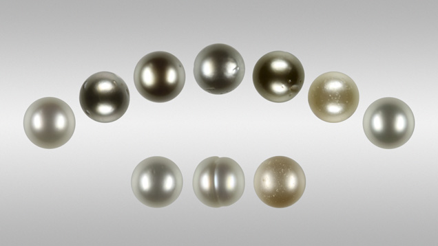
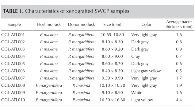
This study was carried out on 10 successfully cultivated experimental SWCPs (selected from McGinty et al., 2010) with various colors and sizes (see figure 1 and table 1). Seven samples (nos. 1–7) were cultivated in P. maxima after transplantation of a P. margaritifera tissue graft, while the other three (nos. 8–10) were cultivated in P. margaritifera after transplantation of a P. maxima graft. All samples were cultivated for 14 months on a farm belonging to Cendanda Indopearls on the Indonesian island of Bali; more on the exact conditions of cultivation can be found in McGinty et al. (2010). None of them had been subjected to any treatment. All but sample 9 were round or near-round, with good to very good surface condition and mostly good luster; see Gübelin Gem Lab (2012) for more information about the grading system used. SWCPs cultivated in P. maxima mollusks with P. margaritifera grafts had more grayish color than those cultivated in P. margaritifera mollusks with P. maxima grafts (again, see table 1).
Karampelas
Figure 1. Ten xenografted saltwater cultured pearls were chosen for this study. Seven samples (nos. 1–7) were cultivated in P. maxima with transplanted P. margaritifera tissue graft, while the other three (nos. 8–10) were cultivated in P. margaritifera with transplanted P. maxima graft. Composite photo by S. Karampelas.
SP13-Karampelas-table1 147466
The samples’ UV fluorescence reaction was observed with a 6W long- and short-wave (365 and 254 nm, respectively) UV lamp. Their UV-Vis-NIR spectra were obtained for the 250–1600 nm range using a Cary 5000 spectrometer fitted with a Varian diffuse reflectance accessory. Only the 250–900 nm range, which contains the color-related absorption bands, is presented here. The data sampling interval and spectral bandwidth of each measurement were set at 0.7 nm and the scan rate at 60 nm/minute. Matte black sample holders were used for a more intense signal. Photoluminescence (PL) spectra were acquired using a Renishaw Raman 1000 spectrometer coupled with a Leica DMLM optical microscope at 50× magnification, with an excitation wavelength of 514 nm emitted by an argon-ion laser (Ar+), a power of 10 mW, a 10-second acquisition time, and a resolution of about 0.1 nm. Digital radiography was performed at the Gübelin Gem Lab with a Comet X-ray unit and a Kodak 6120 digital sensor. Parameters were adjusted to the sample size, with voltage from 60 to 65 kV and current from 5 to 7 mA.

RESULTS AND DISCUSSION

Figures 2–4 show the diffuse reflectance UV-Vis-NIR spectra for six xenotransplanted samples. The spectra present an absorption (a decrease in diffuse reflectance) at around 280 nm. Figure 2 shows two natural-color samples cultivated after xenotransplantation into P. maxima mollusks with P. margaritifera grafts, GGL-ATL002 (dark gray) and GGL-ATL004 (gray), as well as one black natural-color SWCP from P. margaritifera after allotransplantation (bottom spectrum). All three spectra contain six main absorption bands: from 330 to 460 nm, with maxima at 330–385 nm and 385–460 nm, and at 405, 495, 700, and 745 nm (plus a continuous band extending through the visible range with a maximum in the near infrared at around 820 nm). Also observed are three less-intense bands at around 530, 585, and 625 nm, which are common in allotransplanted P. margaritifera SWCPs (Elen 2002; Karampelas et al., 2011). Differences in the spectra patterns are due to the different relative intensities of these bands. The 700 nm band is currently known only from allotransplanted P. margaritifera SWCPs (Elen, 2002). Moreover, the 405 nm band has not been observed in natural-color allotransplanted P. maxima SWCPs (Karampelas, 2012). These results are in accordance with those found experimentally by McGinty et al. (2010 and 2011), as well as other authors (e.g., Wada and Komaru, 1996). In other words, the saibo—in this case, P. margaritifera tissue—is mainly responsible for the coloration of the SWCPs. None of these bands is linked to a specific pigment, except for the one at approximately 405 nm, which is attributed to a kind of porphyrin (Iwahashi and Akamatsu, 1994).
SP13 Karampelas fig2a 147139
Figure 2. These diffuse-reflectance spectra of a black allografted P. margaritifera SWCP (bottom) and two gray xenografted samples from a P. maxima host and P. margaritifera donor (GGL-ATL002 and GGL-ATL004) show absorptions at 280 nm, from 330 to 460 nm (with apparent maxima at 330–385 nm and 385–460 nm), and at 405,495, and 700 nm. Also observed is a weak continuous absorption with a maximum at 820 nm, plus some less-intense features at 530, 585, 625, and 745 nm. For clarity, the spectrum of GGL-ATL004 is shifted up 5% and that of the black natural- color SWCP is shifted down 5%.
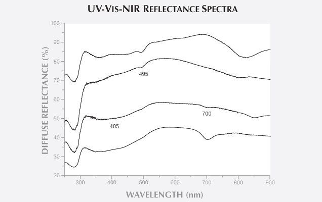
Figure 3 shows the UV-Vis-NIR spectra of two light yellow samples, cultivated after xenotransplantation. Sample GGL-ATL006, cultivated in a P. maxima mollusk with a P. margaritifera graft, is a bit grayish. Sample GGL-ATL010 is cultivated in a P. margaritifera mollusk with a P. maxima graft. Both spectra contain the characteristic absorption feature from 330 to 460 nm observed in yellow to golden natural-color allotransplanted SWCPs from P. margaritifera and P. maxima (Elen, 2002). Both spectra also have a weak band at around 495 nm, similar to yellowish allotransplanted SWCPs of both mollusks (Karampelas, 2012). A weak band at around 700 nm and a shoulder at about 405 nm are also observed in the spectrum of sample GGL-ATL006. These absorption bands, present in allotransplanted SWCPs in P. margaritifera and absent from those cultivated in P. maxima (again, see figure 3), spectroscopically confirm the genetic results from McGinty et al. (2010 and 2011).

SP13 Karampelas fig3a 147455
Figure 3. The light yellow xenografted sample GGL-ATL010 (second spectrum from the top; P. margaritifera host and P. maxima donor) shows a weak absorption feature from 330 to 460 nm (with weak bands at 330–385 nm and 385–460 nm), as well as other bands at 495 nm and in the near-infrared region. Similar bands are observed to analogous natural-color (light brownish yellow) allografted sample from P. maxima (top spectrum), with different relative intensities of the same bands. The light gray yellow xenografted sample GGL-ATL006 (second spectrum from the bottom; P. maxima host and P. margaritifera donor) presents similar absorptions, as well as two additional bands at around 405 and 700 nm. Similar bands are observed in the natural-color allografted sample from P. margaritifera (bottom spectrum). The spectrum for GGL-ATL006 is shifted down 10% for clarity.
Figure 4 presents two samples of very light gray or “white-silver” color from the xenotransplantation of GGL-ATL007 and GGL-ATL008. The two spectra look similar; virtually the entire visible region is transmitted. A weak continuous absorption through the visible range with a maximum in the near-infrared region was responsible for the samples’ light gray color. Very similar spectra can be observed in some white as well as other light-colored (white-silver and light yellow) allografted samples from P. maxima and P. margaritifera. The absorption band at around 700 nm is present in all of the colored samples (allografted or xenografted) cultivated using saibo from P. margaritifera, but was absent from the two light-colored samples (GGL-AUT001 and 007). The 700 nm absorption was absent, or sometimes present as a shoulder, in white to light-colored allotranplanted samples from P. margaritifera (Elen, 2002; Karampelas et al., 2012). Thus, the absence of the 700 nm band from a light-colored SWCP does not preclude the possibility that it was cultivated using saibo from P. margaritifera.


SP13 Karampelas fig4b 147454
Figure 4. Two very light gray (“white-silver”) SWCPs were cultivated after xenografting: GGL-ATL007 (P. maxima “host” and P. margaritifera donor) and GGL-008 (P. margaritifera “host” and P. maxima donor). These display only weak absorptions in the visible region that are not characteristic of either bivalve species. The samples’ very light gray coloration is due to a weak continuous absorption through the visible region, with a maximum in the near-infrared region. The spectrum for GGL-ATL007 is shifted up 10% for clarity.
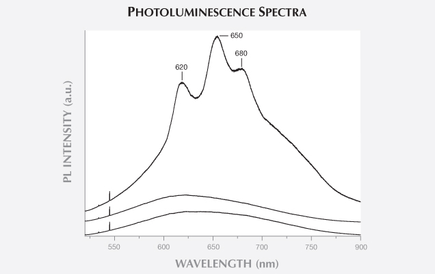
PL spectra of the dark-colored xenografted samples using saibo from P. margaritifera displayed bands in the orange to red region at about 620, 650, and 680 nm with green excitation (figure 5), similar to those in allografted P. margaritifera samples (Miyoshi et al., 1987). The light-colored xenografted samples—cultivated with both grafts—showed less-intense bands (again, see figure 5); similar results were found in allografted samples from both mollusks. Moreover, like allotransplanted SWCPs from the same mollusks, the light-colored samples were inert to short- and long-wave UV radiation (GGL-AUT001 and GGL-AUT006–010), while the others showed a weak greenish yellow and weak yellow reaction, respectively.

SP13 Karampelas fig5 147453
Figure 5. The photoluminescence spectrum of xenografted sample GGL-ATL005 (with saibo from P. margaritifera) shows bands at around 620, 650, and 680 nm. The sharp bands in the 520–550 nm region are due to the Raman effect. The light colored samples (GGL-ATL007 and GGL-ATL008) present less intense bands with a broad apparent maximum around 630 nm. All spectra intensities are adjusted to the main Raman bandand shifted for clarity.
From the X-radiographs, the samples cultivated with a P. maxima donor and a P. margaritifera host generally contained thicker nacre (approximately 1.6– 4.4 mm) than those cultivated using a P. margaritifera donor and a P. maxima host (0.5–1.8 mm; see also table 1). Allografted SWCPs from P. maxima had thicker nacre (as well as nacre weight) than allografted P. margaritifera SWCPs after cultivation for the same period of time in the same farm and under similar conditions; see examples in McGinty et al. (2010). This was probably due to the different growth rate (directly related to the nacre deposition rate) of P. maxima and P. margaritifera bivalves; P. maxima have a higher growth rate than their P. margaritifera counterparts (Yukihira et al., 2006; Saucedo and Southgate, 2008). Nevertheless, the growth rate of P. maxima and P. margaritifera can vary with environmental conditions such as salinity and water temperature (Gervis and Sims, 1992; Yukihira et al., 2006; Saucedo and Southgate, 2008). The radiography results here do confirm that the saibo plays an important role in nacre deposition (McGinty et al., 2010 and 2011).

CONCLUSION

Xenotransplantation between P. margaritifera and P. maxima can yield gem-quality SWCPs, as documented by McGinty et al. (2010 and 2011). This study using UV-Vis-NIR spectroscopy as well as radiography confirmed the histological and genetic findings by various researchers (e.g., Arnaud-Haond et al., 2007; McGinty et al., 2010) that the saibo from the donor mollusk is mainly responsible for the color as well as the nacre thickness. Using spectroscopic means, gemological laboratories can identify (with the exception of some light-colored SWCPs) the mollusk species of the donor (e.g., the 700 nm absorption band characteristic of saibo from P. margaritifera) but not the host. The host mollusk probably plays some role in the nacre deposition. For instance, xenotransplanted SWCPs with a P. margaritifera host and saibo from P. maxima have slightly thicker nacre than the allotransplanted SWCPs from P. maxima (McGinty et al., 2010). Additional research is needed to shed light on this.

Moreover, several studies have shown that selecting the best-secreting saibo for transplantation into a healthy host mollusk is the key to SWCP quality (e.g., Acosta-Salmón et al., 2004; Southgate, 2008). Further research is also needed on all five quality factors in xenografted SWCPs, including comparison with allografted SWCPs from the same mollusk species under identical conditions, after careful selection of donor and host mollusks. These investigations would clearly show if quality can be improved through xenografting. Another meaningful experiment, suggested by various authors, would be to see if xenografting between other Pinctada species (e.g., P. fucata) or even related species (e.g., Pteria sp.) can yield high-quality SWCPs.


Dr. Karampelas (s.karampelas@gubelingemlab.ch) is a research scientist at the Gübelin Gem Lab in Lucerne, Switzerland. Dr. Lombard is a technical manager at Cendanda Indopearls (Atlas South Sea Pearls) in Denpasar, Bali, Indonesia.

Acosta-Salmón H., Martínez-Fernández E., Southgate P.C. (2004) A new approach to pearl oyster broodstock selection: Can saibo donors be used as future broodstock? Aquaculture, Vol. 231, No. 1-4, pp. 205–214, http://dx.doi.org/10.1016/j.aquaculture.2003.08.022.
 
Arnaud-Haond S., Goyard E., Vonau V., Herbaut C., Prou J., Saulnier D. (2007) Pearl formation: persistence of the graft during the entire process of biomineralization. Marine Biotechnology, Vol. 9, No. 1, pp. 113–116 http://dx.doi.org/10.1007/s10126-006-6033-5.
 
Elen S. (2002) Identification of yellow cultured pearls from the black-lipped oyster Pinctada margaritifera. G&G, Vol. 38, No. 1, pp. 66–72, http://dx.doi.org/10.5741/GEMS.38.1.66.

Gervis M.N., Sims N.A. (1992) The biology and culture of pearl oysters (Bivalvia: Pteriidae). International Center for Living Aquatic Resources Management Studies and Reviews, Vol. 21, 49 pp.
 
Gübelin Gem Lab (2012) The Pearl, http://www.gubelingemlab.ch/PDF/GGL_Pearlbooklet_en.pdf [date accessed: Feb. 22, 2013]
 
Haws M. (2002) The Basic Methods of Pearl Farming: A Layman’s Manual. Center for Tropical and Subtropical Aquaculture Publication No. 127, 79 pp., www.ctsa.org/files/publications/CTSA_1276316728619239483681.pdf [date accessed: Nov. 12, 2012].
 
Iwahashi Y., Akamatsu S. (1994) Porphyrin pigment in black-lip pearls and its application to pearl identification. Fisheries Science, Vol. 60, No. 1, pp. 69–71.
 
Karampelas S. (2012) Spectral characteristics of natural-color saltwater cultured pearls from Pinctada maxima. G&G, Vol. 48, No. 3, pp. 193–197, http://dx.doi.org/10.5741/GEMS.48.3.193.
 
Karampelas S., Fritsch E., Gauthier J-P., Hainschwang T. (2011) UV-Vis-NIR reflectance spectroscopy of natural-color saltwater pearls from Pinctada margaritifera. G&G, Vol. 47, No. 1, pp. 31–35, http://dx.doi.org/10.5741/GEMS.47.1.31.
 
Lucas J.S. (2008) Environmental influences. In P.C. Southgate and J.S. Lucas, Ed., The Pearl Oyster. Elsevier, Amsterdam, pp. 187–222.
 
Mamangkey N. (2009) Improving the quality of pearls from Pinctada maxima. PhD thesis, James Cook University (Australia), 167 pp.
 
McGinty E.L., Evans B.S., Taylor J.U.U., Jerry D.R. (2010) Xenografts and pearl production in two pearl oyster species, P. maxima and P. margaritifera: Effect on pearl quality and a key to understanding genetic contribution. Aquaculture, Vol. 302, No. 3–4, pp. 175–181, http://dx.doi.org/10.1016/j.aquaculture.2010.02.023.
 
McGinty E.L., Zenger K.R., Taylor J.U.U., Evans B.S., Jerry D.R. (2011) Diagnostic genetic markers unravel the interplay between host and donor oyster contribution in cultured pearl formation. Aquaculture, Vol. 316, No. 1–4, pp. 20–24, http://dx.doi.org/10.1016/j.aquaculture.2011.02.020.
 
Miyoshi T., Matsuda Y., Komatsu H. (1987) Fluorescence from pearls and shells of black-lip oyster, Pinctada margaritifera, and its contribution to the distinction of mother oysters used in pearl culture. Japanese Journal of Applied Physics, Vol. 26, No. 7, pp. 1069–1072, http://dx.doi.org/10.1143/jjap.26.1069.
 
Saucedo P.E., Southgate P.C. (2008) Reproduction, development and growth. In P.C. Southgate and J.S. Lucas, Ed., The Pearl Oyster. Elsevier, Amsterdam, pp. 137–186.
 
Southgate P.C. (2008) Pearl oyster culture. In P.C. Southgate and J.S. Lucas, Ed., The Pearl Oyster. Elsevier, Amsterdam, pp. 231–272.
 
Tayale A., Gueguen Y., Treguier C., Le Grand J., Cochennec-Laureau N., Montagnani C., Ky C-L. (2012) Evidence of donor effect on cultured pearl quality from a duplicated grafting experiment on Pinctada margaritifera using wild donors. Aquatic Living Resources, Vol. 25, No. 3, pp. 269–280, http://dx.doi.org/10.1051/alr/2012034.
 
Taylor J.J.U., Strack E. (2008) Pearl production. In P.C. Southgate and J.S. Lucas, Ed., The Pearl Oyster. Elsevier, Amsterdam, pp. 273–302.
 
Wada K., Komaru A. (1996) Color and weight of pearls produced by grafting the mantle tissue from a selected population for white shell color of the Japanese pearl oyster Pinctada fucata martensii (Dunker). Aquaculture, Vol. 142, No. 1–2, pp. 25 – 32, http://dx.doi.org/10.1016/0044-8486(95)01242-7
 
Yukihira H., Lucas J.S., Klumpp D.W. (2006) The pearl oysters, Pinctada maxima and P. margaritifera, respond in different ways to culture in dissimilar environments. Aquaculture, Vol. 252, No. 2–4, pp. 208–224, http://dx.doi.org/10.1016/j.aquaculture.2005.06.032.