The First Identified Non-Nacreous Beaded Cultured Pearl

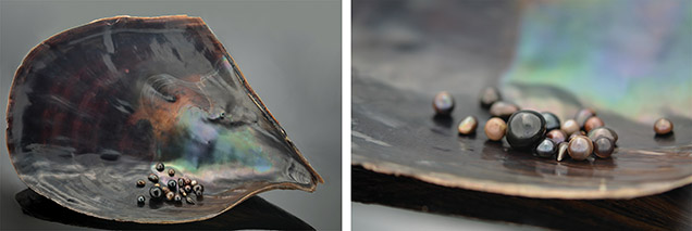
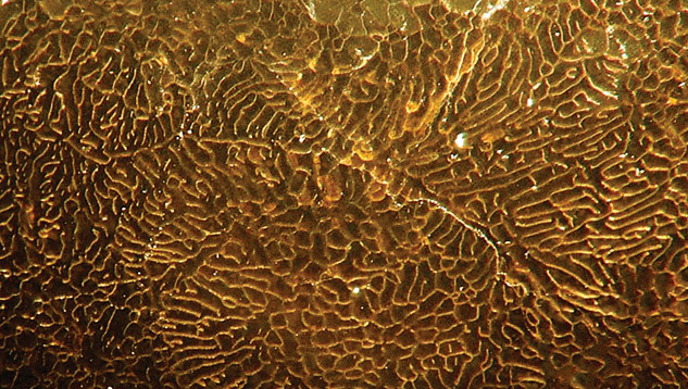
Its dark black porcelaneous appearance was typical of pearls from the calcitic part of Atrina vexillum, the kind usually submitted for laboratory analysis (figure 1). Observation at 85× magnification (figure 2) revealed a distinctive pattern of convolution, with a few micro-cracks.

Figure 2. Surface magnification of the non-nacreous beaded cultured pearl revealed a typical calcitic pattern of pen pearls. Photo by Olivier Segura; magnified 85×.
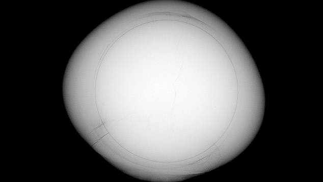
Our findings on these cultured pearls are similar to those of N. Sturman et al. (“Observations on pearls reportedly from the Pinnidae family [pen pearls],” Fall 2014 G&G, pp. 202–215) in terms of Raman spectra (confirming the calcitic nature), UV-Vis spectra, and chemical composition.The most interesting findings came from X-radiography. Microradiography/tomography imaging clearly shows a bright white disk inside the pearl (figure 3). The continuous separation line around this feature leaves no doubt that there is a bead inside the cultured pearl. As the beads used to nucleate “nacreous” pearls typically come from large freshwater mussels, we know that they are of a slightly denser material than saltwater nacre. Thus, they appear whiter than the surrounding material (of saltwater origin). The bead’s appearance was consistent with a classical nucleus of the kind used in bead-nucleated pearls. Measurement of the bead gave a 6.70 mm diameter.

Figure 3. This microradiograph clearly identifies the pen pearl as a beaded cultured pearl. Image by Olivier Segura.
Tomography confirmed the presence of a near-perfect spherical bead inside the cultured pearl. Even with the owner’s consent, however, we decided not to cut the pearl, as the imaging techniques were sufficient to prove the bead-cultured origin.We wonder if this gem represents a single specimen achieved by a curious experimenter, or if there is now an industrialized process for cultivating such pearls. The cultivation technique used, even if it is known to produce cultured pearls in farms, requires advanced knowledge of the process. The owner reports having handled a black round pen pearl “the size of a golf ball.” From the size and symmetry and given this new discovery of a bead-cultured pen pearl, one can speculate that this too might be a bead cultured pearl.
This bead nucleation technique for unusual species could have easily been developed in the past. To our knowledge, though, this specimen represents the first non-nacreous beaded cultured pearl examined by a laboratory. Faced with the expansion of the bead culturing process and the possibility of successful trials in different mollusks, gemologists need to be very cautious in natural origin determinations, even when there is every reason to believe a pearl to be natural.
.jpg)


