The Grass Is Always Greener…
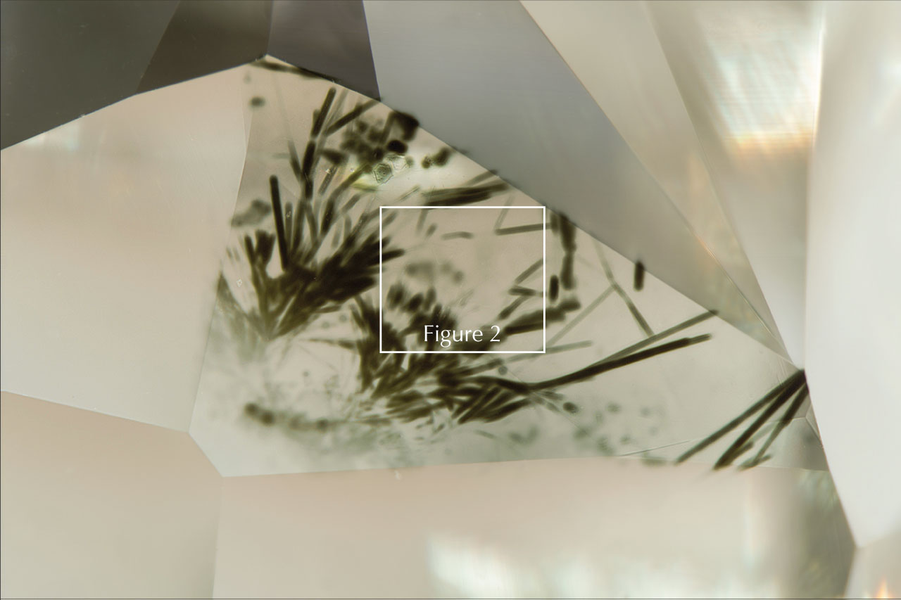
The Carlsbad laboratory recently examined a 0.97 ct Fancy grayish green diamond submitted for colored diamond grading service. Microscopic examination revealed a colorless diamond with green radiation stains scattered across the pavilion (figure 1).
These radiation stains were particularly unusual due to their linear appearance and resembled blades of grass in their morphology and color. These stains were prevalent along intentionally preserved rough natural diamond surfaces on the pavilion. Faceted surfaces along the pavilion were polished with particular effort to retain some of the green radiation stains. The resultant internal reflection of green light to the table created a uniform green bodycolor in this rare and beautiful diamond.
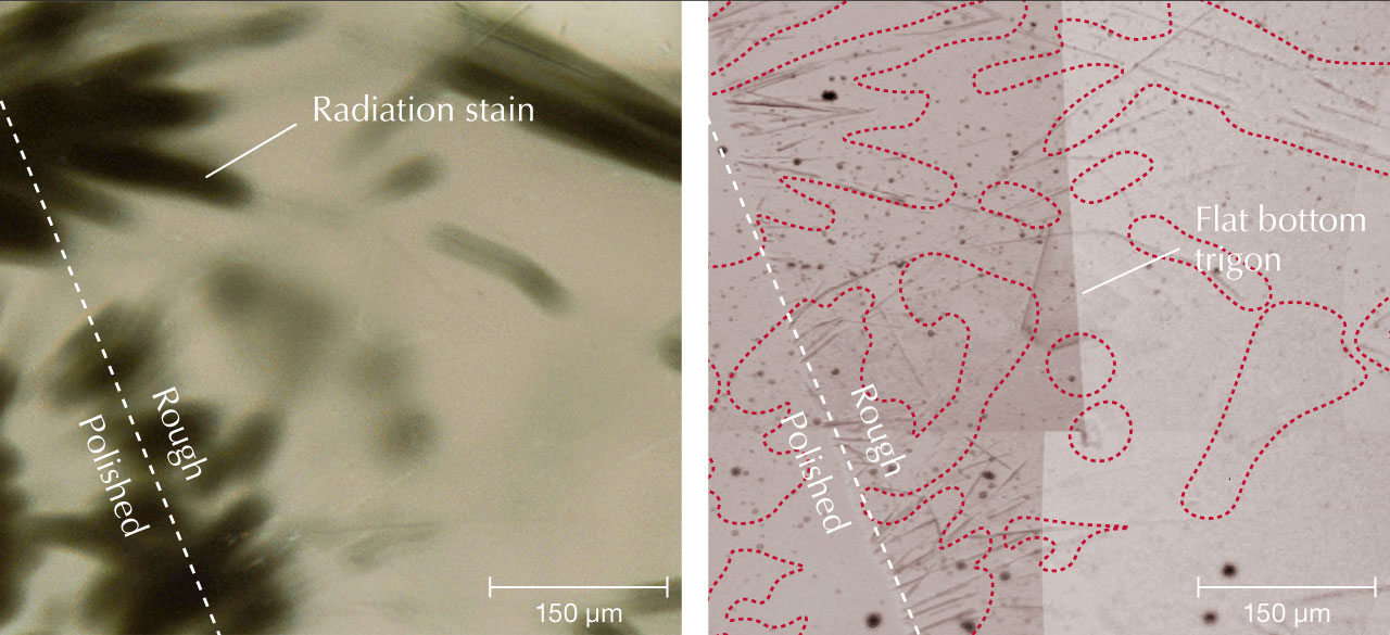
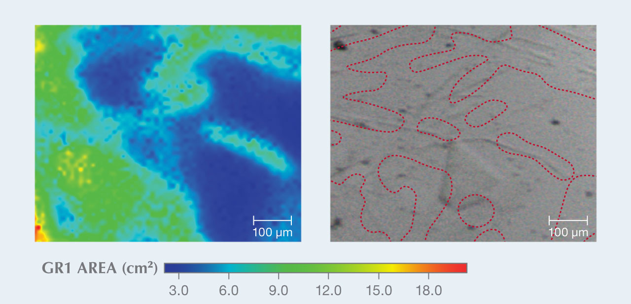
Surface photomicrographs revealed long, linear, and acicular indentations concentrated near the center of the radiation stains (figure 2). Indentations crosscut flat-bottomed trigons, suggesting they post-dated diamond dissolution in the earth’s mantle (e.g., R. Tappert and M. Tappert, Diamonds in Nature: A Guide to Rough Diamonds, Springer, Heidelberg, 2011). The green stains along the polished surface indicate that the radiation stains penetrated some depth into the diamond. These features have been observed previously at GIA (e.g., Spring 2021 G&G Micro-World, pp. 66–67; Spring 2022 G&G Micro-World, pp. 64–65) and may result from acicular to tabular radioactive minerals precipitating directly on the diamond surface in a paleoplacer to recent alluvial environment (e.g., R. Lauf, Mineralogy of Uranium and Thorium, Schiffer Publishing, Ltd., Atglen, Pennsylvania, 2016). Radiation damage created vacancies in the diamond lattice, which absorb red light and reflect green light, resulting in the green color of the radiation stains. Uncharged or neutral vacancies produce a photoluminescence response near the 741 nm zero phonon line called the GR1 defect (e.g., J.E. Shigley and C.M. Breeding, “Optical defects in diamond: A quick reference chart,” Summer 2013 G&G, pp. 107–111). The GR1 defect area from photoluminescence spectra can be measured and mapped to indicate the spatial distribution of this defect coincident with the radiation stains (figure 3).
It is unusual to see green radiation stains of this shape that correspond with indentations on a diamond surface. These features could be a commonly overlooked characteristic of green diamonds.


