Two Foraminifera-Like Objects Found in a Natural Saltwater Pearl

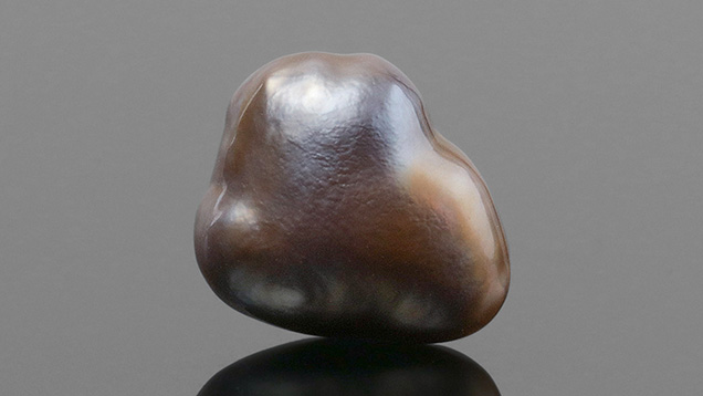
GIA’s Hong Kong laboratory recently received a brown nacreous pearl weighing 1.57 ct and measuring 11.06 × 9.15 × 7.20 mm (figure 1). It had a baroque shape and exhibited a soft luster with an unaltered surface. Despite its size, the pearl felt relatively lightweight, indicating the possibility of a hollow or partially hollow internal structure. Viewed under 40× magnification, the surface displayed overlapping nacre platelets in a spiral pattern, similar to that observed in pearls produced by Pteria species mollusks (L. Kiefert et al., “Cultured pearls from the Gulf of California, Mexico,” Spring 2004 G&G, pp. 26–39).
The sample exhibited the typical brownish surface tint of pearls produced by the Pteria mollusk species. Its ultraviolet/visible spectrum showed the characteristic reflectance features of naturally colored pearls formed in this mollusk, with identifiable absorptions at 405 and 495 nm (S. Karampelas et al., “Spectral differentiation of natural-color saltwater cultured pearls from Pinctada margaritifera and Pteria sterna,” Summer 2011 G&G, p. 117). Under long-wave UV radiation, the pearl also displayed moderate red fluorescence, a reaction linked to a type of porphyrin pigment found in pearls originating from the Pteria species (Kiefert et al., 2004). Energy-dispersive X-ray fluorescence analysis showed no traces of manganese and 1150 ppm of strontium, confirming its saltwater origin.

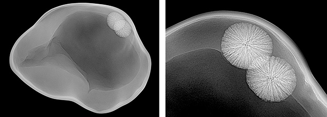
Real-time microradiography revealed a fascinating internal structure (figure 2). A large central void, composed of light and dark gray areas associated with the presence of organic matter, occupied almost the entire interior of the pearl, which explained the lighter than expected heft. Surrounding the void, a few distinct growth arcs were observed, following the outline of the pearl’s shape—a typical feature seen in natural pearls. In addition, two intriguing foreign materials were trapped within the inner wall of the light gray organic-rich area of the void.

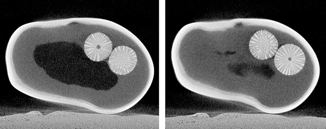
Further analysis via X-ray computed microtomography (μ-CT) imaging revealed the porous nature of these two materials. Resembling foraminifera, a marine micro-skeleton member of a phylum of amoeboid protists, they consisted of radial structures extending from the center of empty cores (figure 3). The pair of near-spherical objects measured 1.50 × 1.25 mm and 1.53 × 1.33 mm, respectively, and appeared to be separate entities while sharing a homogeneous formation. The pearl’s structure was judged to be natural due to the appearance of the natural-looking void, which consisted of a flowy outline, and the presence of the foraminifera-like entities. It is worth noting that most cultured pearls from the Pteria species available in the market are bead cultured pearls. Non-bead cultured “keshi” Pteria pearls are normally of smaller sizes and distinguishable internal structures.
GIA has received numerous pearl submissions in the past with interesting internal structures related to foreign materials. In fact, the skeletal composition of the foreign material observed looked exceedingly similar to that found in a natural pearl examined by GIA’s Bangkok laboratory in 2015 (Winter 2015 Lab Notes, pp. 434–435). However, this pearl stands out for the presence of paired foraminifera-like spheres. No two pearls are the same, and it is always rewarding when advanced testing reveals such captivating features.
.jpg)


