A Natural Pearl with an Intriguing Internal Structure

Aspects of natural pearl formation remain a mystery in many cases; however, one generally accepted principle is that an intruder finds its way into the mantle or gill areas of a mollusk and instigates the pearl formation process. Gemologists are never sure what they will find when natural pearls are examined using microradiography. Substantial real-time X-ray microradiography (RTX) work by GIA staff in various locations has shown evidence of formation cause in only a few pearls.
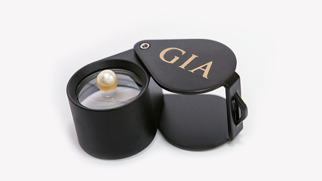
Early in 2015, a cream semi-baroque button pearl measuring 5.22 × 5.04 mm and weighing 0.95 ct (figure 1) was submitted to GIA’s Bangkok laboratory with nine other pearls. Externally the pearl appeared similar to many other previously examined natural pearls, but the microradiography uncovered something that was far from normal.
After initial RTX examination, the apparent initiator of this specimen’s formation was found to be very different from anything encountered, to our knowledge, in a natural pearl. RTX revealed an attractive radiating structure within a central conchiolin-rich boundary and associated external arcs within the surrounding nacreous overgrowth (figure 2). The radial object displayed clear solid arms extending from the central core of the structure. Given the nature of the core, further investigation was performed in order to identify the radial feature and see whether the pearl was natural or formed via human intervention using an organic nucleus as a “bead.”
Computed microtomography (μ-CT) analysis was used to perform a 3-D examination of the radial feature. The μ-CT results revealed a magnificent structure with a snowflake-like intricacy to its form (figure 3). The structure created some doubts about the natural formation of the pearl. The dark organic/conchiolin-rich growth structure, with some gaps between the nucleus and the surrounding nacre, has been noted in other unusual natural pearls of known origin. This differs from the so-called “atypical bead-cultured pearls” that almost always display a tight structure (lacking growth arcs) around the inserted nucleus. Almost all of the whole shell nuclei experimentally inserted into Pinctada maxima hosts have shown the boundary of the shell/nacre interface to merge tightly with one another, with very little in the way of organic arcs within the surrounding nacre. This curious structure led to further investigations into published claims of natural organic nuclei within natural pearls.
Similar but not identical structures have been documented in RTX and μ-CT work carried out on two pearls recovered from natural P. maxima shells by GIA staff (“An expert’s journey into the world of Australian pearling,” 2014). Pearls with unique structures have also been reported by others (K. Scarratt et al., “Natural pearls from Australian Pinctada maxima,” Winter 2012 G&G, pp. 236–261). The exact origin of the pearls studied in Scarratt et al. is unclear, although they are more likely to be natural in at least two of the examples cited. For instance, the whole shell of one sample mentioned by Scarratt et al. showed characteristics similar to the specimen described here, with visible arcs within the nacre surrounding a shell nucleus that has a notable void/organic interface between the shell and nacre. The very small drill hole present in the shell, as opposed to the larger drill holes commonly found in cultured pearls, also strongly suggests a natural origin.
The nucleus of the 0.95 ct pearl under discussion appeared to be an organism such as coral. In order to find a match, coral samples were analyzed by RTX and μ-CT methods to compare their structures. Results indicated that the feature within the pearl may well be coral, but no exact match was noted. During the recent International Gemmological Conference (IGC) in Vilnius, Lithuania, it was suggested that the similar internal feature of a different pearl (N. Sturman et. al, “X-ray computed microtomography (μ-CT) structures of known natural and non-bead cultured Pinctada maxima pearls,” Proceedings of 34th International Gemmological Conference, 2015, pp. 121–124) could well be a remarkably preserved foraminifera, a marine micro-skeleton member of a phylum of amoeboid protists. They are usually less than 1 mm in size; their structures vary, and many live on the sea floor. Given the environment, size, and features evident in the form within the pearl under discussion, it may well be that the internal structure is a type of foraminifera sphere with a porous structure for passive filtration (see figure 6 at www.mdpi.com/1660-3397/12/5/2877/html). The μ-CT results revealed the interconnecting channels and radial structure characteristic of these spherical foraminifera.
Whatever the true nature of the central form, the author considers the structure of this pearl to be natural based on the characteristics noted. It is also interesting to see that this pearl was small and not of particularly fine quality, other characteristics of natural pearl samples collected from the field. GIA’s own experiments with small pieces of shell and coral using P. maxima hosts have produced larger pearls with different external appearances and generally finer quality than the specimen studied here. Further research into the differences between unusual structures in natural and atypical bead-cultured pearls will continue.


