Inclusions and Spectroscopic Features of Yellowish Green Enstatite

Gem-quality enstatite is the magnesium end member of the enstatite-ferrosilite series in the clinopyroxene subgroup of the pyroxene group. While the most common color is brown, it can also be colorless, green, or gray. Myanmar, Tanzania, and Sri Lanka are the main sources of gem-quality stones currently in the market. Enstatite can yield clean faceted gems but can also display four-, six-, or eight-rayed asterism.
Enstatites have been documented many times from various sources. Six-rayed star brown enstatite was reported from India (W. Eppler, “Star-diopside and star-enstatite,” Journal of Gemmology, Vol. 10, No. 6, 1967, pp. 185–188). Sri Lankan samples were reported in U. Henn and H. Bank, “Sternbronzit aus Sri Lanka,” Zeitschrift der Deutschen Gemmologischen Gesellschaft, Vol. 40, No. 2–3, pp. 145–148. Star enstatite from Madagascar was described in T. Cathelineau, “Six-rayed star enstatite from Madagascar,” Journal of Gemmology, Vol. 36, No. 8, 2019, pp. 688–690. Four-rayed star brown enstatites from Sri Lanka were documented in E.J. Gübelin and J.I. Koivula, Photoatlas of Inclusions in Gemstones, Volume 3, Opinio Publishers, Basel, Switzerland, 2008. Faceted brown enstatite was reported in Koivula et al., “Gemmological investigation of a large faceted East African enstatite,” Journal of Gemmology, Vol. 21, No. 2, 1988, pp. 92–94. Yellowish green enstatites from Africa were discussed in K. Schmetzer and H. Krupp, “Enstatite from Mairimba Hill, Kenya,” Journal of Gemmology, Vol. 18, No. 2, 1982, pp. 118–120, as well as in B.M. Laurs et al., “Yellowish green enstatite (and star enstatite) from Tanzania,” Journal of Gemmology, Vol. 36, No. 8, 2019, pp. 691–693. Norwegian stones were documented in F. Schmitz et al., “Polymer-filled star enstatite from Norway,” Journal of Gemmology, Vol. 35, No. 2, 2016, pp. 98–101.
Recently, GIT Gem Testing Laboratory (GIT-GTL) received six bright yellowish green enstatite samples weighing 0.65 to 2.78 ct (figure 1) that were reported by author SD to be from Africa. The stones appeared fairly similar to the previously documented four-rayed star yellowish green enstatite from Tanzania documented by Laurs et al. (2019). In that article, needle-like inclusions were found to be the cause of the four-rayed asterism, but their mineralogical identity was not reported.
Of the six stones examined in this study, four were cabochons displaying a four-rayed star effect while two were faceted (figure 1). The five stones with at least one polished flat surface gave refractive indices of α = 1.655–1.660, β = 1.661–1.665, and γ = 1.668–1.669, with a birefringence of 0.009–0.014, and are therefore biaxial positive. The spot RI reading of one cabochon without a flat surface was ~1.66. The hydrostatic specific gravity of all samples was 3.22–3.25. All these values fall well within the range of enstatite. The stones appeared weak orange under the Chelsea color filter and showed moderate yellow and yellowish green pleochroism. All were inert to both long-wave and short-wave UV.
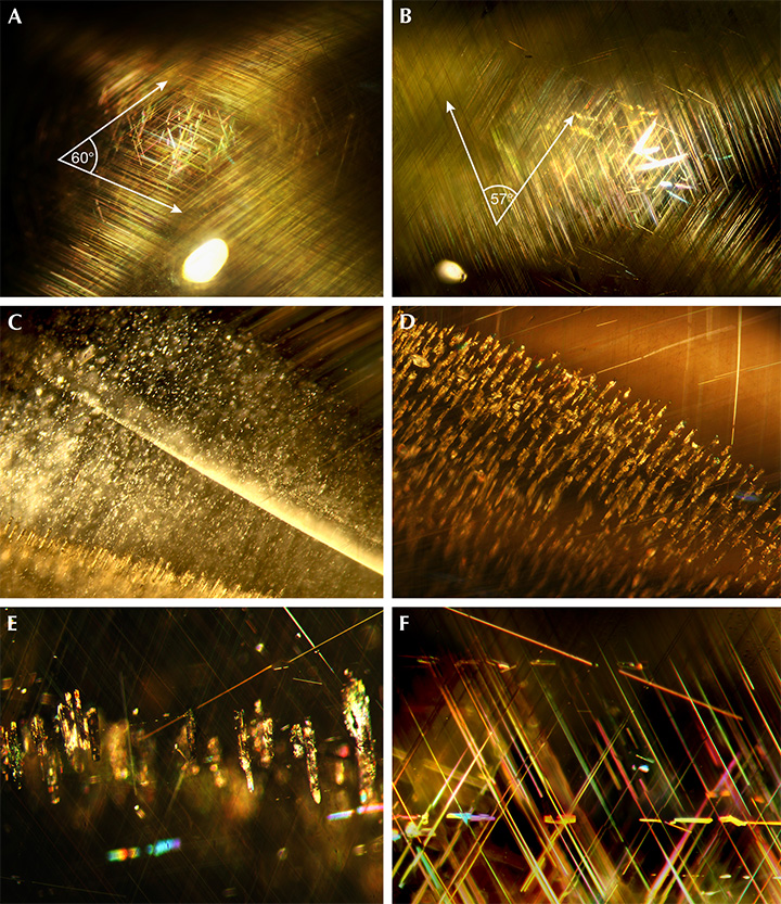
The main diagnostic internal features were the two sets of dense and regularly oriented needle-like inclusions (figure 2, A and B). The two sets measured on the same plane at the top of three cabochons (e278, e132, and e144) were found to intersect at an approximate angle of 57° to 60°, producing the four-rayed star effect seen on the cabochon surface (figure 1). While the two faceted stones also contained fine needle-like inclusions, they were not cut to display a star.

Close examination also uncovered two sets of needles that did not produce asterism. In addition, patches of densely oriented minute white particles (figure 2C) and a plane of oriented short tubes (figure 2, D and E) were found in one stone (e144). These tubes were partially filled with tiny brownish white to colorless crystals identified by Raman spectroscopy as magnesite (figure 3). The presence of epigenetic magnesite (MgCO3) could have resulted from alteration of the host enstatite. The other feature consisted of sparse rectangular platelet inclusions occurring in various directions within the dense mesh of needle inclusions (e067, figure 2F).
The Raman spectra of all host mineral samples, recorded using 532 nm laser excitation, similarly showed dominant peaks that matched the enstatite reference spectrum from the RRUFF database (not shown here). Moreover, the Raman spectrum of the exposed fine needle inclusions plus host mineral in one sample (e078) gave the superimposed peaks of rutile (440 and 610 cm–1) and enstatite (238, 343, 662, 685, 1010, and 1030 cm–1), consistent with the RRUFF reference spectra in figure 3. Similarly, the Raman spectrum of exposed white to colorless crystals in short tubes plus the host in a sample (e144) showed overlapping peaks of magnesite (213, 330, 737, and 1094 cm–1) and enstatite, also consistent with the RRUFF reference spectra. The identity of the rectangular platelet inclusions, however, could not be resolved due to their sparsity and their depth below the surface.

Polarized ultraviolet/visible/near-infrared (UV-Vis-NIR) spectra of a sample (e132) showed features dominated by broad absorption bands of Fe2+ in the NIR region centered at ~900 and ~1750 nm (figure 4). The spectra rise continuously from ~700 nm toward the UV with small Fe2+ peaks at 427, 506, and 546 nm in the visible range (Cathelineau, 2019). In addition, these iron absorption features were superimposed by absorption bands around 420–490 nm and 600–700 nm due to the presence of V3+ with trace Cr3+ (see table 1). These two bands cause the absorption of the violet-blue and orange-red ends of the visible spectrum. Such overlapping absorption features allow the transmission window in the green to the yellow region, giving rise to the bright yellowish green color (Laurs et al., 2019). The origin of the absorption band at ~2220 nm, however, could not be resolved.

Semi-quantitative EDXRF chemical analyses revealed enriched contents of silicon (58.1–59.2 wt.% SiO2), magnesium (26.5–31.5 wt.% MgO), and aluminum (6.3–8.1 wt.% Al2O3), with trace amounts of iron, calcium, titanium, vanadium, chromium, manganese, and zinc (table 1). Furthermore, the end-member compositions were calculated and appeared close to the enstatite end members (92.0–96.2% enstatite, 3.5–7.5% ferrosilite, and 0.3–0.5% wollastonite) of the enstatite-ferrosilite series.

In summary, the attractive bright yellowish green colors of these enstatites are caused by the combination of significant iron and vanadium with a trace of chromium substituting magnesium in the crystal lattice. The four-rayed asterism phenomenon is caused by light reflection from two sets of very dense fine rutile needle inclusions intersecting one another at approximately 57° to 60°. The similarity of these stones and those reported by Laurs et al. (2019) (tables 1 and 2) leads us to conclude these stones could also be from Tanzania.


