Natural Diamond with Unusual Phosphorescence

Phosphorescence spectroscopy shows that natural type IIb diamonds can have two broad phosphorescence bands: one centered at 500 nm that appears greenish blue and one centered at 660 nm that appears red. Generally, one of these phosphorescence bands is dominant. In the Hope diamond, for example, the phosphorescence initially appears orangy red and transitions to red as the subordinate greenish blue phosphorescence fades away (S. Eaton-Magaña et al., “Using phosphorescence as a fingerprint for the Hope and other blue diamonds,” Geology, Vol. 36, No. 1, 2008, pp. 83–86). Occasionally, the faster-decaying greenish blue phosphorescence band will transition to the slower-decaying red band (Spring 2007 Lab Notes, pp. 47–48).
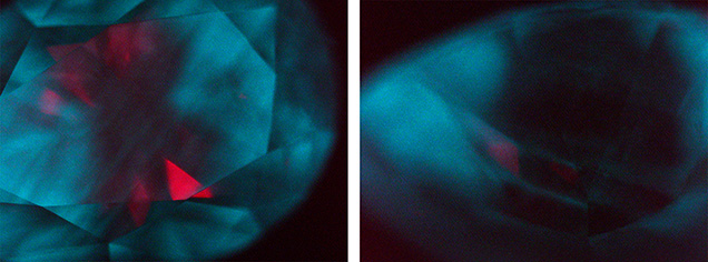
Recently, a 0.90 ct diamond with D color and I1 clarity was submitted to GIA’s laboratory in Ramat Gan, Israel, for a diamond dossier report. Infrared (IR) absorption spectroscopy indicated that the natural diamond was nominally type IIa. However, photoluminescence (PL) spectroscopy revealed that the diamond contained boron-related features, including the 648.2 nm peak attributed to a boron interstitial (B. Green, “Optical and magnetic resonance studies of point defects in single crystal diamond,” PhD thesis, University of Warwick, 2013), along with the observed phosphorescence. These features indicated that the diamond did contain boron, but below the detection level of IR absorption spectroscopy. The distinctive aspect was that both the red and greenish blue phosphorescence were localized and visible within the same image (see above).
The phosphorescence is attributed to donor-acceptor pair recombination. For the 500 nm band, this recombination has been identified as interactions between boron acceptors and isolated nitrogen donors (J. Zhao, “Fluorescence, phosphorescence, thermoluminescence and charge transfer in synthetic diamond,” PhD thesis, University of Warwick, 2022), while the 660 nm band is attributed to boron acceptors and unknown donors that are likely related to plastic deformation (S. Eaton-Magaña and R. Lu, “Phosphorescence in type IIb diamonds,” Diamond and Related Materials, Vol. 20, No. 7, 2011, pp. 983–989). PL maps were collected on the table and pavilion with 455, 532, and 785 nm excitation; only an undefined PL feature at 813 nm showed an apparent increase on the pavilion within the red-phosphorescing region (see above, right).
Type IIb diamonds grown by high-pressure, high-temperature (HPHT) methods almost always show the 500 nm phosphorescence and occasionally a separate orange phosphorescence band centered at 580 nm. In a few instances, HPHT-grown diamonds have shown the 500 and 580 nm bands simultaneously in different portions of the diamond (Eaton-Magaña and Lu, 2011; Spring 2023 Gem News International, pp. 155–156).
This is the first time the authors have seen spatially distinct observations of red and greenish blue phosphorescence in a natural diamond, an unusual find.


