Non-Bead-Cultured Pearls from Pinctada margaritifera
April 27, 2018
INTRODUCTION
Gray to black pearls from the Pinctada margaritifera (black-lipped pearl oyster) bivalves are commonly referred to in the market as “Tahitian pearls” (figure 1). This name is almost always associated with cultured pearls (beaded and non-beaded). While gray to black are the most likely colors encountered, these pearls do exhibit a range that extends from light tones all the way to dark tones of gray to black. When their sometimes dramatic overtones—or mixes of other secondary hues—are included in the cocktail, their appearances can change dramatically, producing some very striking pearls that are in global demand. These overtones and/or secondary hues may be green, pink, purple, or a combination of colors leading to such exotic and descriptive terms as “Peacock,” “Aubergine,” or “Pistachio.” On rare occasions, white, brown, and yellow primary bodycolors with a range of overtones may also be found.
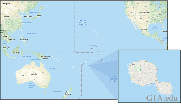
The Pinctada margaritifera mollusk’s habitat extends over a wide range but is most often associated with the Indo-Pacific region (Strack, 2006). The species almost always shows characteristic gray to black coloration over the mother-of-pearl that comprises the bulk of its body and which in turn is responsible via the genetics of its mantle for producing the recognizable color of Tahitian pearls. Nowadays the majority of Tahitian pearls produced commercially originate from French Polynesia (figure 2), a group of islands located in the south-central Pacific Ocean. Tahiti is the largest of the islands within the group and lends its name to the type of pearl under discussion.
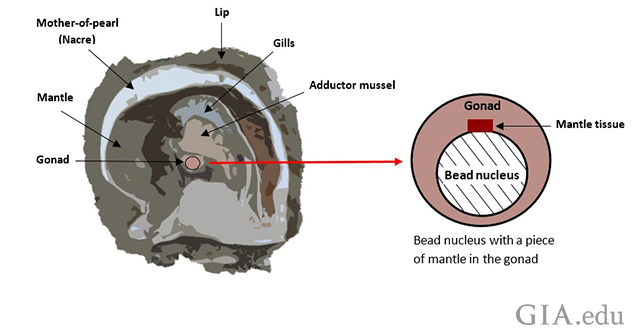
The process used to produce cultured pearls involves operating on the mollusks and nurturing them while the pearls develop within the shells. The usual technique employed in creating a bead-cultured (BC) pearl is to surgically insert a freshwater bead nucleus (the majority of nuclei are fashioned from freshwater shell) into the gonad of a host mollusk with a small piece of mantle tissue from a donor mollusk (figure 3). The mantle tissue is responsible for secreting the nacre that in turn covers the bead nucleus and determines the color of the pearl (Goebel, 1989). Should the bead be rejected from the pearl sac for any reason, the result may be the potential formation (though not in every case) of an accidental non-bead-cultured pearl (NBC), beadless cultured pearl, or “keshi” pearl that results from the piece of mantle tissue that was inserted or from cells from the piece of mantle that may have separated. There are other possibilities. If the pearl sac already exists, and this operation is the second (or beyond) where just a bead is inserted and no mantle is necessary, an NBC may also be formed if the bead is rejected (Hänni, 2006).
The majority of pearls obtained from the farms of the French Polynesian islands are BC pearls, though not all the nuclei inserted produce such a pearl and thus some NBC pearls also result as a by-product. This type of pearl is valuable and in demand in its own right, so they are not always unwelcome and create another product that can be marketed by the trade (Doulton, 2015). Since little has been published in the gemological literature about the internal structures of NBC pearls produced by the Pinctada margaritifera mollusk, GIA took the opportunity to obtain a selection of cultured pearls on loan from a dealer in Bangkok to study their structures as well as their spectral features for reference purposes. Only by examining the structures of pearls of known types from known sources can any reliable database be developed. While we were unable to source the pearls in this study directly from the farm/mollusk, we believe we obtained pretty reliable samples from a trusted source who handles cultured pearls on a daily basis. The pearls were also selected by GIA team members on the premises and removed from bags of similar-looking pearls numbering in the thousands. The chances of any truly natural pearls being included among the pearls would be slim at best. The results of the analysis appear to support the claims that these pearls were cultured in Pinctada margaritifera bivalves, or at least were formed from mantle that incorporated the genes of the Pinctada margaritifera mollusk (Karampelas, 2013). The various internal structures recorded and the characteristic spectra that relate to the various colors examined will provide valuable references for gemologists looking for data on such NBC pearls.
MATERIALS AND METHODS
A total of 120 samples, reported to be Tahitian cultured pearls, were examined for this study. The source from whom we acquired the samples claimed they were all NBC pearls from various farms within French Polynesia. Groups A (68 pearls) and B (47 pearls) were hand-picked at random from two large bags of keshi pearls on the premises of Orient Pearl Company in Bangkok by representatives of GIA Bangkok’s pearl team, who visited the factory during August 2016. The five pearls in Group C were handed to the team separately when they asked to see the largest NBC pearls available (figure 4).

Pearls from Groups A and B measured between 7.68 × 7.32 × 5.68 mm and 16.25 × 7.84 × 7.64 mm and weighed from 0.59 to 6.49 ct, while those from Group C ranged from 16.40 × 14.38 × 14.11 mm to 18.40 × 16.95 × 13.80 mm and from 19.95 to 29.50 ct. The pearls were of various shapes and possessed a range of colors from silver to black. Some samples exhibited more than one color.
The internal structures of the pearls were examined using real-time microradiography (RTX) and X-ray computed microtomography (μ-CT). The overall group microradiographic images were obtained using a real-time Matrix-FocalSpot XT-3 X-ray inspection system with a 5-micron microfocus, a voltage of 90 kV and a current of 0.18 mA, X-ray source, and a dual 4"/2" image intensifier (II) with digital camera. The more detailed individual microradiographic images were obtained via a Pacific Industries (PXI) GenX-90 X-ray system with a 4-micron microfocus, 90 kV voltage and 0.18 mA current X-ray source, and a PerkinElmer 4"/2" dual-view flat panel detector. A ProCon CT-mini X-ray system with a 5-micron microfocus, 90 kV voltage and 0.18 mA X-ray current source and a Hamamatsu flat panel detector with 50 μm pixel pitch and a frame grabber card were used to obtain all the μ-CT data.
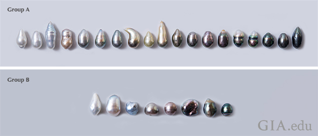
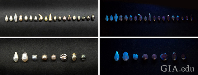
Twenty-seven (27) pearls of various colors were selected from Groups A and B and sorted by tone and color intensity (light to dark of various hues) in order to study the characteristic spectra of each type for reference and to make sure they were all naturally colored. The samples chosen from Group A (19 pearls) and Group B (8 pearls) were photographed to show their tones and hues/saturations (figure 5). Their ultraviolet and visible spectral features were recorded using an Agilent Cary 60 UV-Vis spectrophotometer with a custom-made GIA integrated sphere accessory incorporating an 80 Hz xenon flash lamp source. Spectra were recorded in the 200–800 nm range with a data interval set at 1.0 nm and a 1.5 nm fixed spectral bandwidth and scan rate of 184.62 nm/min. Raman and photoluminescence (PL) spectra were obtained using a Renishaw inVia reflex micro-Raman spectrometer system with a 514 nm argon-ion laser at 50 mW excitation. The laser was set at 100% power with three accumulations and an exposure time of 10 seconds. PL spectra were collected with the laser power set at 50% with one accumulation and an exposure time of 15 seconds.
The fluorescence reactions of the pearls were also observed upon exposure to ultraviolet (UV) radiation and ultra-short-wave radiation. For these tests we used a GIA-designed long-wave UV unit with a 4-watt UV lamp emitting 365 nm radiation and a DTC DiamondView unit with a xenon flash lamp source and a special ultraviolet filter that produces short-wavelength ultraviolet radiation of less than 225 nm.
Once sawn in half, photomicrographs of the pearls' interior features were taken using a Nikon SMZ18 stereomicroscope with SHR Plan Apo 1× objective lens and NIS-Elements imaging software.
INTERNAL STRUCTURES
All the pearls were examined by real-time microradiography (RTX) to reveal their internal structures. As expected, most of them revealed void and/or “linear” features associated with organic-rich areas typical of most NBC pearls (Sturman, 2009). However, five BC pearls were found mixed with the samples, while some of the other samples revealed questionable or ambiguous structures that needed more detailed µ-CT analysis.
The samples from Groups A and B were sorted into smaller groups for RTX analysis owing to the limited field of view (FOV) of the RTX machine. Thus, only a general overview of the structures could be obtained prior to examining the individual NBC pearls in more detail. The group RTX results revealed various types of void and linear structures (table 1) and also identified two pearls from Group A and three pearls from Group B as BC pearls that required no further X-ray analysis.
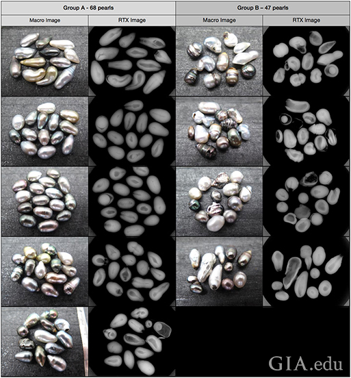
appearance shown on the left and their internal structures on the right.
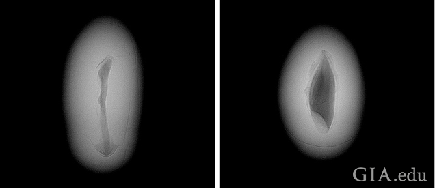
The detailed microradiographic work on the pearls in Group A revealed that the majority possessed void structures, some with organic-rich areas surrounding the voids. The void forms varied slightly from the thinner, elongated void feature in Pearl A-12 to the broader and more obvious void feature in Pearl A-23 (figure 6). These voids are characteristic of saltwater NBC pearls from Pinctada species mollusks.
Besides the usual void features, other variations were also observed in some pearls. The descriptions that follow show their differences. The structures of the pearls are shown in table 2 in the sequence in which they will be discussed in further detail, from Pearl A-15 to A-66.
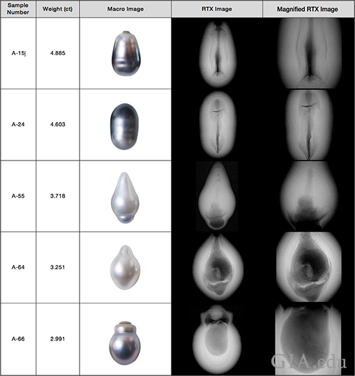
Pearl A-15 (figure 7) revealed an elongated void with a clear organic-rich area around the structure that ran the length of the pearl. The void actually reached the surface at one end, and on first impression the pearl appeared to be partially drilled, though it was not. The overall form of the structure and appearance of the pearl were enough to conclude that it was an NBC pearl.
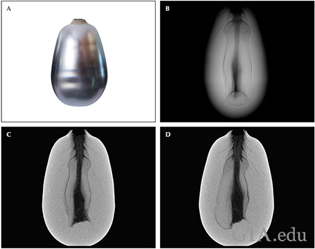
of its internal structure (B). Two µ-CT slices reveal the internal details more clearly (C, D).
Pearl A-24 (figure 8) showed an elongated void structure with some short cracks branching out at one end of the pearl. An organic-rich “meaty” area surrounds the void feature. Cracks such as these may be observed in NBC and natural pearls, though there are some subtle differences.
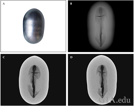
of its internal structure (B). Two µ-CT slices reveal the internal details more clearly (C, D).
Pearl A-55 (figure 9) showed a fairly large void with some organic matter (a gray feature in the thicker lower area of the RTX image). The void extends upward toward the tapered end and narrows in the central area. Distinct growth arcs may be seen extending from the lower void feature and are also clearly seen following the pearl’s shape toward the tapered end. The structure of this pearl is not as convincing as the others in table 2, but its overall form and external appearance indicate it is more likely an NBC pearl than a natural pearl. GIA would interpret such a structure as NBC.
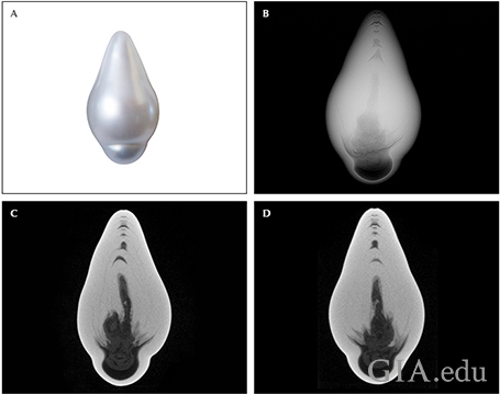
of its internal structure (B). Two µ-CT slices reveal the internal details more clearly (C, D).
Pearl A-64 (figure 10) revealed a very clear void with a small white feature possessing a short linear structure at its center. Some organic matter (gray features) is also visible within the void. This type of void, with small solid white features inside, is most often associated with NBC pearls. The linear structure within the small white object further supports an NBC formation.
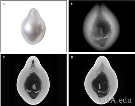
of its internal structure (B). Two µ-CT slices reveal the internal details more clearly (C, D).
Pearl A-66 (figure 11) showed a more evenly outlined void structure near the center with a “meaty” organic structure around it. Very little structure is present within the void, just one “solid” feature at the bottom that extends upward slightly. This is probably an organic “fold/wall.” This structure is characteristic of saltwater NBC pearls. The upper “twin” growth area of the pearl and its underlying structure are not so helpful when it comes to separating NBC pearls from natural pearls, but the pearl’s overall appearance and the main void in the primary pearl growth are rather diagnostic.
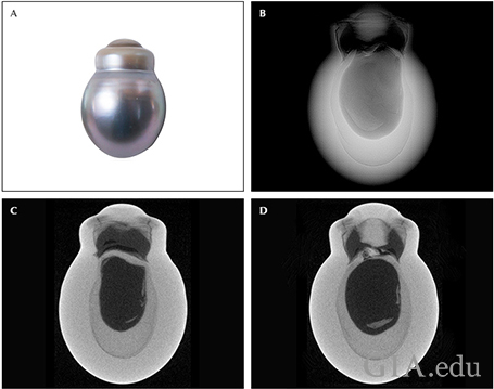
of its internal structure (B). Two µ-CT images reveal the internal details more clearly (C, D).
The microradiographic results of the pearls in Group B also revealed voids as their predominant features (table 1, right). However, some showed linear structures and others revealed structures that would be challenging to gemologists examining the pearls on a “blind” basis (submitted without any information on origin/history) under strict laboratory conditions. Pearls B-13 and B-21 (figure 12) are examples of the linear and void structures observed within two of the NBC pearls studies in the group, respectively.
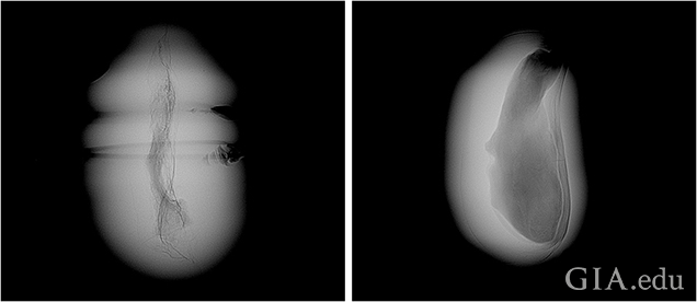
RTX images of some of the more interesting structures encountered in pearls from Group B, together with their weights, are included in table 3 and discussed in more detail below the table with additional µ-CT data for further clarity.
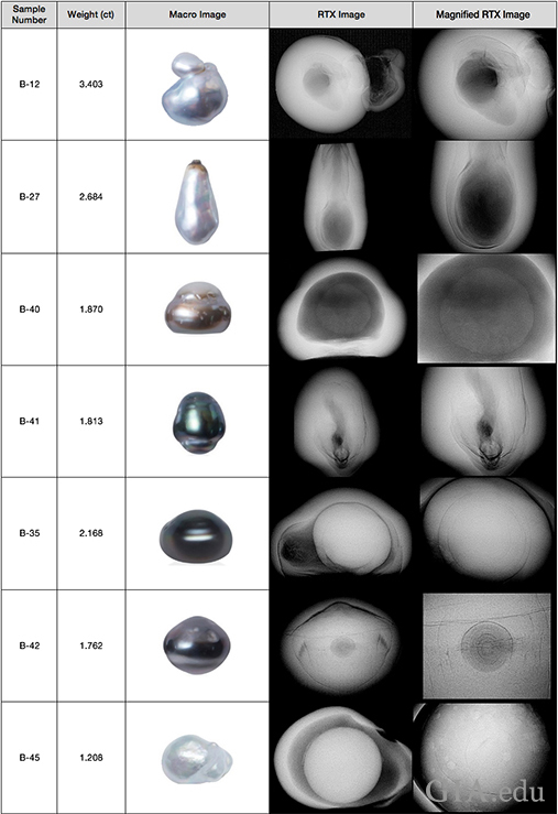
when examined by RTX analysis.
Pearl B-12 (figure 13) showed a “double structure” consisting of two voids, one in each part of the twin -type pearl. The main (lower) part exhibited an irregular void in the center with a faint “meaty” organic-rich halo. The smaller attached Tokki-type feature also revealed an irregular void, but was relatively much larger and contained some structural walls. µ-CT imaging revealed the details more distinctly with the walls showing as an abundance of tiny white particles. The main evidence for the sample’s NBC origin is the form of the void in the main part of the pearl, which is fairly characteristic of such pearls.
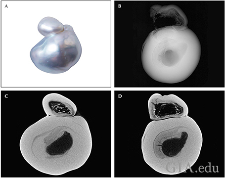
of its internal structure (B). Two µ-CT images reveal the internal details more clearly (C, D).
Pearl B-27 (figure 14) showed a fairly large void in the thickest area of the drop that extended up toward the tapered end, together with a faint organic-rich meaty area that surrounded the void. Clear growth arcs may be seen curving downward from the tapered end through the meaty area. This pearl’s structure shares some similarities (the tapering form of the void and the apparent partially drilled nature) with that seen in Pearl A-15. However, Pearl B-27 is much closer to the structures seen in some natural pearls and may be considered natural by some gemologists. Given its association and origin, it would be very unlikely that the pearl is in fact natural, so a strong case can be made for NBC.
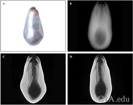
of its internal structure (B). Two µ-CT images reveal the internal details more clearly (C, D).
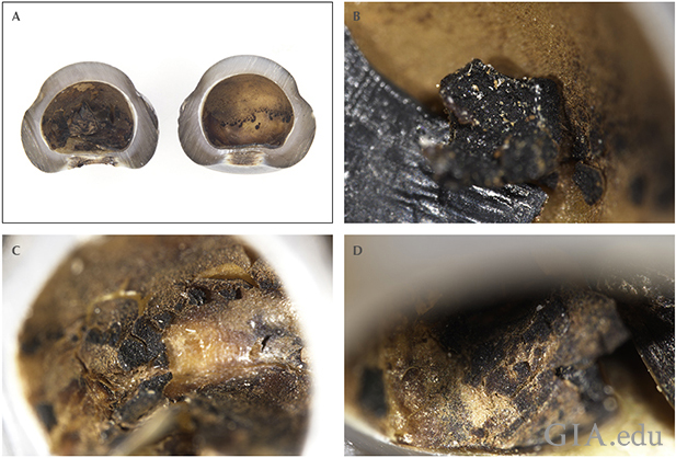
Pearl B-40 (figure 15) revealed a large, fairly smooth void with a more radio-opaque (though not as radio-opaque as the pearl body) inhomogeneous gray object within it. The object did not take the form of a bead nucleus or anything that could be considered “atypical” (Hänni, 2010; Scarratt, 2017) and intentionally introduced into the mollusk to create a pearl. Hence, it is more likely of organic nature but with a form not often seen in NBC pearls. Since the owner was convinced the pearl was cultured, permission was given to cut the pearl in half in order to see the interior clearly. Microscopy showed that the saw blade, together with the water used as a coolant, must have shattered or dissolved most of the object as only a few minute pieces remained. These pieces did not seem to possess any obvious conchiolin characteristics, so they were likely “mud” or some other organic matter since Raman analysis also failed to narrow down the identification. Given the likely organic and non-crystalline structure observed under a gemological microscope, this did not come as any surprise. The fact that the piece disintegrated/dissolved so readily also makes it less likely it was conchiolin, based on the authors’ experience. The majority of what remained was more typical of areas of conchiolin, and these may be seen in figure 16.
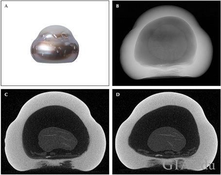
of its internal structure (B). Two µ-CT images reveal the internal details more clearly (C, D).
Pearl B-41 (figure 17) showed a small white “seed”-like feature in some orientations that resolved into a much longer oval form in others. In all orientations we noted a short linear feature at its core, similar to that seen in the white feature of sample A-64, and the seed itself is positioned within a void area that leads to a meaty organic area. The void structure with the linear-centered white seed within it is the main structure that proves this pearl is a NBC pearl. The external appearance of this pearl was also more characteristic of an NBC pearl.
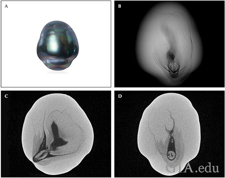
of its internal structure (B). Two µ-CT images reveal the internal details more clearly (C, D).
While all the pearls shown in the tables and discussed to this point may possess structures that the majority of pearl-testing gemologists would consider NBC, others examined in this study revealed structures that would no doubt lead to lengthy discussions and possible disagreements on their identity if submitted blind to a gemological laboratory. For example, Pearl B-35 (figure 18) looks rather suspect externally, and this would immediately lead to doubts about any natural formation, even though nothing internally would strike a gemologist as being characteristic of a NBC origin. There is an obvious outline separating a solid white “pearl” exhibiting nice growth arcs from the core to the outer layers, from a void feature to one side. There is also a meaty area following the boundary. The only concerns would be the weak white structures within the void, the meaty boundary and obvious lack of clear structure surrounding the meaty structure, but they are hardly sufficient evidence to identify the pearl as NBC. We also considered the possibility of it being an “atypical bead-cultured pearl” (aBC), but insufficient evidence was observed. While suspicious, this pearl does not really possess enough features to prove whether it is cultured pearl or natural, and it would most likely have to be considered “undetermined.”
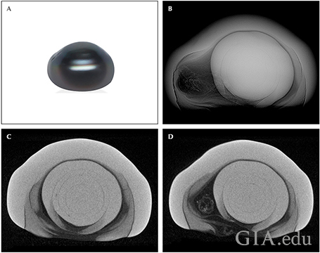
of its internal structure (B). Two µ-CT images reveal the internal details more clearly (C, D).
Pearl B-42 (figure 19) also showed an ambiguous structure. While the external appearance once again seemed to indicate a cultured origin, the small dark concentric core with a white circular center is certainly more characteristic of natural origin than cultured. The associated surrounding growth arcs and lack of anything that would even remotely indicate an NBC origin all point toward natural. Once again, if submitted on a blind basis, there is no logical reason why this pearl would not receive a “natural pearl” result on a gemological report. The only very minor feature that may raise a red flag is some tiny white seeds (one indicated on µ-CT slice “c”) within the most prominent growth arc visible. However, this is not enough evidence to identify the pearl as NBC.
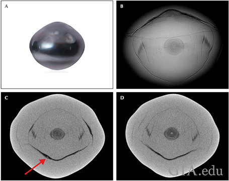
of its internal structure (B). Two µ-CT images reveal the internal details more clearly (C, D).
The minute "seed" that may indicate a cultured origin is shown, but the evidence is too
subjective to be considered as proof. All the evidence points to a natural classification.
In keeping with a previous pearl (B-35), another externally suspect sample, Pearl B-45 (figure 20), showed a very clear inner solid feature within a large irregular void. Both pearls have what appears to be a separate body (“pearl”) within a “host” pearl, although it is much more obvious in this example. The inner pearl in each exhibits a clear concentric structure typical of natural pearls, and there is nothing to indicate a cultured origin other than the possibility that the inner pearls have been intentionally inserted into an existing pearl sac to produce an atypical BC pearl. While this may be a possibility, the pearls do not appear to be aBC examples, and the owner was adamant that no such culturing takes place at any of the farms from which the samples were sourced. Thus, they are more likely to be NBC or natural. The patchy features within the void area share some similarities with the voids seen in known NBC samples previously studied by GIA (Sturman, 2016), but the overall structures of the pearls are still not enough to prove they are NBC. As such, this pearl would also likely be considered “undetermined.”
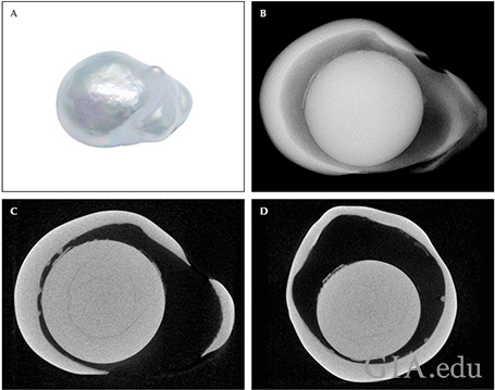
of its internal structure (B). Two µ-CT images reveal the internal details more clearly (C, D).
As the last three samples (B-35, B-42 and B-45) in particular prove, some pearls from this research showed questionable internal structures that may be challenging to identify as NBC pearls. While the source and the information available on their origin would lead us to believe they are more likely NBC, the fact remains that if they were submitted to a gemological laboratory for identification on a blind basis, disagreements on their true identifies may result. These differences of opinion can happen not only between different laboratories but also between staff of the same laboratory. Such are the challenges faced by pearl testers from time to time.
The five large pearls in the final group (Group C), were all examined by RTX and µ-CT analysis to reveal their internal structures. The RTX images and their weights are shown in table 4 and the pearls are presented in the order discussed below (C-1 to C-5). Although three of them showed characteristic NBC structures, the two remaining pearls revealed more intriguing features.
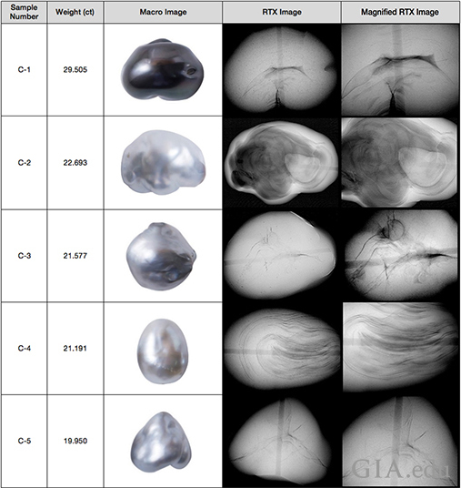
Pearl C-1 (figure 21) revealed a narrow curved void structure in the center with some weak whitish features inside. The associated arcs appeared to flow around the void, and overall the structure is characteristic of saltwater NBC pearls. The external appearance also appeared consistent with NBC pearls from the Pinctada margaritifera mollusk, and the similarity to the majority of those already discussed should be noted.
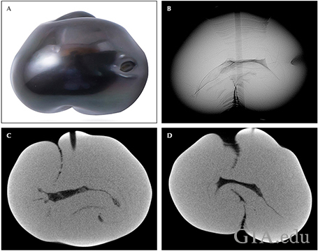
structure (B). Two µ-CT images reveal the internal details more clearly (C, D).
Pearl C-2 (figure 22) showed a large irregular void structure, but the latter did not look completely empty as clear weaker circular features were also visible, as was a solid white object with a central linear structure to one end where the partial drill hole is positioned. The circular features are likely organic matter, similar to that observed and described for Pearl B-40. Overall, this pearl’s features and external appearance are all characteristic of an NBC pearl. Nonetheless, it was very interesting to note the delicate and intricate yet subtle pattern within the void, which was probably made clearer by the likely expulsion and/or drying of any fluids or evacuation of any gas once the pearl was partially drilled.
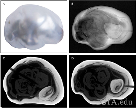
of its internal structure (B). Two µ-CT images reveal the internal details more clearly (C, D).
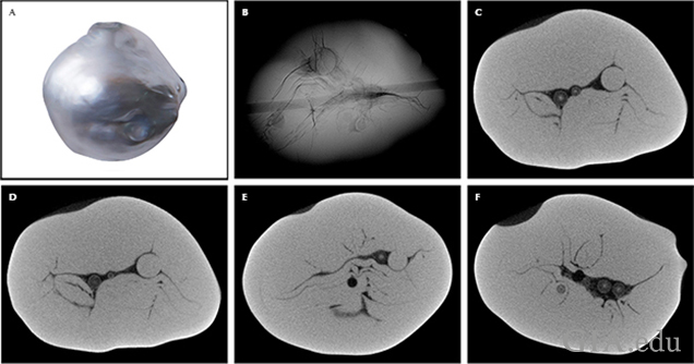
Pearl C-4 (figure 24) revealed the most challenging structure of the five largest pearls, although one that would likely be considered NBC by experienced pearl-testing gemologists. It consisted of a series of very clear concentric rings of an odd shape that formed around a central feature that resembled a void in some orientations and was less obvious in others. The µ-CT images showed the details more clearly, and the overall interpretation led us to believe that this was an NBC pearl with structure that could be somewhat misleading to some pearl testers with limited experience.
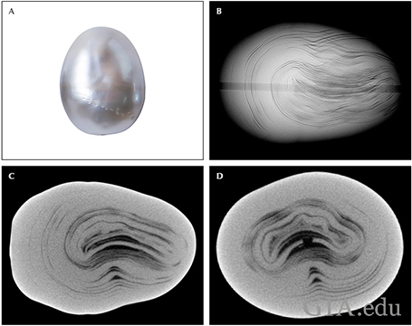
internal structure (B). Two µ-CT images reveal the internal details more clearly (C, D).
Pearl C-5 (figure 25) showed a characteristic NBC “Y-shaped” linear structure at its center with a few minor linear features branching out from it. In addition, a small circular seed feature with surrounding concentric growth rings was observed next to one arm of the linear feature. Although the concentric structure and the form of this paticular seed often indicates that a pearl is more likely to be natural, this was not the case with this pearl, and once again the external appearance as well as the predominantly linear structure are sufficient to conclude this was an NBC pearl.
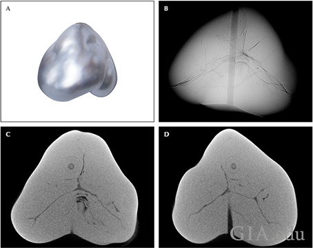
of its internal structure (B). Two µ-CT images reveal more clearly internal structure (C, D).
SPECTROSCOPIC RESULTS
Some low-quality saltwater and freshwater pearls may be treated to imitate the colors seen in Tahitian pearls. The identification of their color origin is necessary to separate them from the naturally colored samples. By studying the UV-Vis, Raman and PL spectra of known naturally colored Tahitian cultured pearls, or at least of reportedly known natural coloration, a useful reference database can be created. Previous research and related articles have shown that pearls produced by the Pinctada margaritifera species exhibit characteristic spectroscopic features that conclusively identify their natural color origin and prove they originated from the mollusk species.
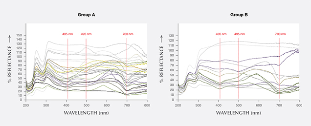
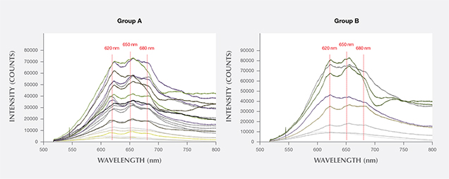
The characteristic spectra recorded for natural-color Pinctada margaritifera pearls include a “bumpy” or “uneven” appearance in the Raman data; three peaks in the PL spectra at approximately 620, 650, and 680 nm; and three features in the UV-Vis spectra around 405, 495, and 700 nm (Elen, 2002; Karampelas, 2011). It is the 700 nm feature in the visible region of the UV-Vis spectrum that narrows the mollusk down to Pinctada margaritifera or potentially Pinctada mazatlanica (Homkrajae, 2016). The reflectance features observed in the UV-Vis spectra around 405 nm and the PL features at approximately 620 nm, usually with others around 650 and 680 nm, are reportedly associated with the protein uroporphyrin, a color pigment usually found in pearls from the black-lipped oyster (Miyoshi, 1987; Iwahashi, 1994).
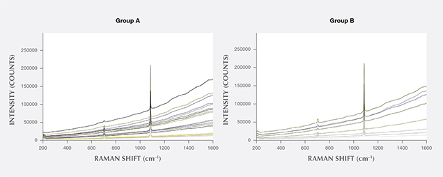
Spectroscopic analyses of the samples in this study (table 5) employed the three methods discussed above and are shown by colored spectra in the figures. The spectral features can be seen to vary in accordance with the tone and saturation of the pearl sample. The darker and more saturated the color, the darker the corresponding spectrum in the diagrams. Almost all the pearls examined showed typical Pinctada margaritifera features, with the lighter-colored pearls showing weaker features than the more saturated and darker samples. Some of the white to light gray samples did not exhibit the expected features, but this is not entirely surprising since they are almost pigment-free or at least have pigment concentrations below those necessary to reveal clear spectral features.
The UV-Vis spectra obtained showed different features in the visible range from 400 to 700 nm, and all the spectra showed the feature at approximately 280 nm in the ultraviolet region. Pearls with the lightest colors, white to light gray, almost all showed “flat” visible spectra owing to the lack of any pigments or their weaker pigment concentrations. This is in keeping with the majority of white pearls. Exceptions may exist, though, as Pearls A-46 and A-2 prove (figure 26 and table 5). Both showed weak features around 405 and 495 nm, while Pearl A-2 also revealed a feature at 700 nm. The light brown and light yellow samples showed features around 405, 495 and 700 nm that varied in intensity with the tone and saturation of the samples. The gray, brown, purplish gray, and purplish brown pearls revealed the three features above more clearly and the darkest, most saturated samples (dark gray to black) showed the most obvious features in the same positions. The features at 405, 495, and 700 nm are all characteristic of naturally colored Pinctada margaritifera or Pinctada mazatlanica pearls, but since we were assured that the samples were sourced from various farms within French Polynesia, we can be reasonably confident that they are in fact Pinctada margaritifera pearls.
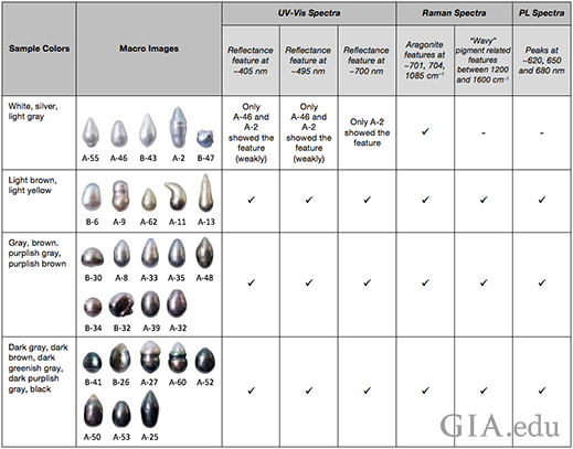
The results of the Raman analysis revealed obvious aragonite peaks at 701/704 (doublet), and 1085 cm–1, proving their mineral composition (Urmos, 1991), while features between 1200 to 1600 cm–1 showed features corresponding to their color origin. The white to light gray samples produced smooth, relatively flat featureless spectra in this region. The light brown and light yellow samples showed weak wavy spectra, whereas the gray, brown, purplish gray, and purplish brown samples had more defined wavy spectra. However, the darkest and most saturated pearls, dark gray to black, produced spectra with the most distinct wavy features (figure 27 and table 5).
Likewise, the PL spectra obtained revealed that the lightest-color pearls, white to light gray, displayed smoother almost featureless spectra in the region of interest. The light brown and light yellow samples revealed weak features around 620, 650, and 680 nm similar to those observed in previous research on Tahitian pearls (Elen, 2002; Karampelas, 2011). The gray, brown, purplish gray, and purplish brown pearls showed clearer features around 620, 560, and 680 nm, while the darkest and most saturated pearls, dark gray to black, produced the most obvious spectral features (figure 28 and table 5), consistent with the results obtained for the UV-Vis and Raman spectra.
The long-wave ultraviolet (LWUV) fluorescence reactions were also observed for interest (figure 29). The results clearly showed different reactions relative to the tones and saturations of their bodycolors. The lightest-color samples, white to light gray, exhibited a strong bluish green fluorescence. The light brown and light yellow samples displayed more muted reactions of a moderate bluish green, but any lighter areas on them exhibited a strong bluish green reaction similar to the one seen on the white and light gray samples. The gray, brown, purplish gray, and purplish brown pearl samples showed very weak to weak bluish green fluorescence, while the darkest and most saturated samples—again dark gray to black—were inert.
The final test we carried out was to observe the fluorescence reactions of some of the samples in the DiamondView. All twenty-seven (27) samples exhibited a blue reaction over most of their surfaces (figure 30, A and B), with moderate to strong intensity. Some variation, weak to strong blue, was noted in samples possessing uneven or circled surfaces (figure 30C). An organic-rich area on one sample with a non-nacreous surface showed an atypical strong greenish yellow reaction (figure 30D). It is interesting to note that the general moderate to strong blue fluorescence is not influenced by the hue, tone, and saturation in the same way as the results obtained by the other instruments.
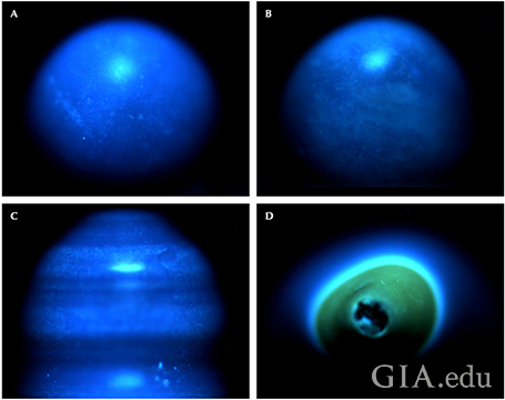
Pearl A-35 (A) and Pearl B-30 (B) exhibited a fairly homogeneous blue fluorescence,
while Pearl A-27 (C) showed some differences in intensity owing to the circled nature of
the surface. Pearl A-50 (D) showed a strong greenish yellow reaction at one end where
the structure was organic rich and non-nacreous, but the rest of the nacreous pearl also
showed a strong blue fluorescence, in keeping with the majority of samples.
CONCLUSIONS
The majority of Pinctada margaritifera NBC pearl samples in this work revealed obvious internal structures during RTX examination. Some others revealed more interesting and ambiguous structures that required further µ-CT analysis to help in their identification. The RTX and µ-CT structures of the 120 samples were saved for future reference. Only five pearls were an exception to the claimed NBC identity of the samples, and all five revealed a clear bead nucleus, proving their BC status. Externally, the “new” appearance of all the samples was similar, so RTX was the only certain way of separating them from the rest of the pearls, which shows how easy it is to accidentally mix pearls of different types when harvesting. Only 3% of the samples showed questionable structures—those that would prove challenging to conclusively identify in normal laboratory testing conditions. Potential doubts about these pearls could lead to an “undetermined” result or even inaccurate results since laboratory gemologists are only able to identify pearls from what they see and by comparing, if necessary, samples against any known references available at the time of submission. Since the authors were informed that all the pearls originated from pearl farms, they should in theory all be classified as cultured (BC or NBC). However, the evidence obtained for some did not prove their NBC identity beyond a doubt, thus proving how challenging pearl testing can be in practice.
Pinctada margaritifera pearls are known to exhibit characteristic UV-Vis spectra that enable both the species and natural color origin to be identified with a high degree of confidence. The Raman and PL spectra provide further supporting evidence for mollusk and color origin determination. The majority of the samples studied here showed characteristic spectral features usually associated with Pinctada margaritifera pearls, which suggests that they likely formed within the mollusk or were at least genetically formed using Pinctada margaritifera mantle during the culturing process. The lightly colored white to light gray samples hardly showed any characteristic features, though a couple of them displayed weak UV-Vis features on areas that likely contained more pigment (slightly darker areas). Even though the features were apparent, they were too weak to assist in the mollusk origin and so were of no practical use in confirming in which mollusk they formed. Since Pinctada mazatlanica pearls may show spectral features similar to those shown by Pinctada margaritifera pearls (Homkrajae, 2016), it is not always straightforward to separate pearls produced by these two mollusks. However, when pearls under examination show characteristic cultured structures, as did the majority of the samples tested in this study, their origin is almost certainly linked to Pinctada margaritifera since this mollusk is the only one of the two that produces cultured pearls on a commercial scale.
Ms. Nilpetploy (née Somsa-ard) is a staff gemologist, and Ms. Lawanwong and Mr. Kessrapong are analytics technicians, in pearl services at GIA in Bangkok.
The authors would like to thank Mr. Devchand Chodhry, managing director of Orient Pearl Company in Bangkok, for providing the samples examined in this study. Nuttapol Kitdee, digital image technician at GIA in Bangkok, is also thanked for his assistance with photography. Appreciation is also extended to all the other GIA staff members who assisted in this work.
Doulton M. (2015) Keshi pearls: Gorgeous flukes of nature. The Jewellery Editor, Jul. 5, http://www.thejewelleryeditor.com/jewellery/article/keshi-pearls-gorgeous-flukes-nature/
Elen S. (2002) Identification of yellow cultured pearls from the black-lipped oyster Pinctada margaritifera. Gems & Gemology, Vol. 38, No. 1, pp. 66–72, https://www.gia.edu/gems-gemology/spring-2002-yellow-cultured-pearls-pinctada-margaritifera-elen
Goebel M., Dirlam D.M. (1989) Polynesian black pearls. Gems & Gemology, Vol. 25, No. 3, pp. 130–148, https://www.gia.edu/gems-gemology/fall-1989-black-pearls-goebel
Hänni H.A. (2006) A short review of the use of 'keshi' as a term to describe pearls. The Journal of Gemmology, Vol. 30, No. 1/2, pp. 51–58.
Hänni H.A., Krzemnicki M.S., Cartier L. (2010) Appearance of new bead material in cultured pearls. The Journal of Gemmology, Vol. 32, No. 1–4, pp. 31–37.
Homkrajae A. (2016) Gem News International: Spectral characteristic of Pinctada mazatlanica and Pinctada margaritifera pearl oyster species. Gems & Gemology, Vol. 52, No. 2, pp. 207–208.
Iwahashi Y., Akamatsu S. (1994) Porphyrin pigment in black-lip pearls and its application to pearl identification. Fisheries Science, Vol. 60, No. 1, pp. 69–71.
Karampelas S., Lombard A. (2013) Gemological characteristics of saltwater cultured pearls produced after xenotransplantation. Gems & Gemology, Vol. 49, No. 1, pp. 36–40, https://www.gia.edu/gems-gemology/spring-2013-karampelas-cultured-pearls-xenotransplantation
Karampelas S., Fritsch E., Gauthier J.P., Hainschwang T. (2011) UV-Vis-NIR reflectance spectroscopy of natural-color saltwater cultured pearls from Pinctada margaritifera. Gems & Gemology, Vol. 47, No. 1, pp. 31–35, https://www.gia.edu/gems-gemology/spring-2011-saltwater-pearls-karampelas
Miyoshi T., Matsuda Y., Komatsu H. (1987) Fluorescence from pearls and shells of black lip oyster, Pinctada margaritifera, and its contribution to the distinction of mother oyster in pearl culture. Japanese Journal of Applied Physics, Vol. 26, No. 7, pp. 1069–1072.
Scarratt K., Sturman N., Tawfeeq A., Bracher P., Bracher M., Homkrajae A., Manustrong A., Somsa-ard N., Zhou C. (2017) Atypical "beading" in the production of cultured pearls from Australian Pinctada maxima. GIA Research News, https://www.gia.edu/gia-news-research/atypical-beading-production-cultured-pearls-australian-pinctada-maxima
Strack E. (2006) Pearls. Rühle-Diebener, Stuttgart, Germany.
Sturman N. (2008) Pearls with unpleasant odors. Lab Notes, http://www.giathai.net/pearls-with-unpleasant-odors/
Sturman N. (2009) The microradiographic structures of non-bead cultured Pearls. Lab Notes, http://www.giathai.net/microradiographic-structures-cultured-pearls/
Sturman N., Bergman J., Poli J., Homkrajae A., Manustrong A., Somsa-ard N. (2016) Bead-cultured and non-bead-cultured pearls from Lombok, Indonesia. Gems & Gemology, Vol. 52, No. 3, pp. 288–297, https://www.gia.edu/gems-gemology/fall-2016-bead-cultured-pearls-lombok-indonesia
Sturman N., Manustrong A., Pardieu V. (2016) The cultured pearls of Mergui with an emphasis on their internal structures. Proceedings of the 5th GIT International Gem and Jewelry Conference, Pattaya, Thailand, pp. 143–145.
Urmos J., Sharma K., Mackenzie F.T. (1991) Characteristic of some biogenic carbonates with Raman spectroscopy. American Mineralogist, Vol. 76, pp. 641–646.
.jpg)


