Unmasking Emerald Filler

Of the treatments we see in the laboratory, fissure filling has become one of the most ubiquitous. We have observed this treatment in a variety of stones, including emerald, ruby, sapphire, spinel, tourmaline, tanzanite, and more. The filling of fractures minimizes their appearance, making the gems appear cleaner. Fissure filling can be detected with a variety of methods. One way is to examine the infrared spectrum, where some fillers display distinctive peaks. Another is the hot point method, where oils can be observed leaking out in droplets. Some fillers are unmasked with simple observation in the microscope, because they display flashes of color or because they include visible dyes.
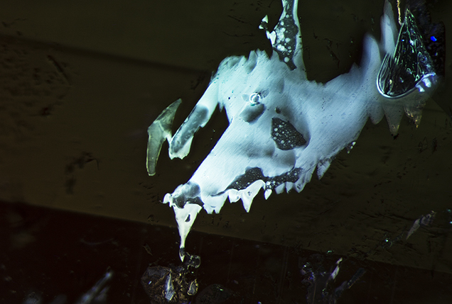
In addition to these methods, another tool in our arsenal is the long-wave ultraviolet flashlight, which can be used in conjunction with the microscope. Because some fillers display a chalky fluorescence when illuminated with long-wave UV light, shining a long-wave UV flashlight at the specimen is a simple technique that helps us to not only to detect the filler, but also to see its exact location in the stone. This helps the gemologist gauge the extent of filling and its impact on the stone’s overall appearance.

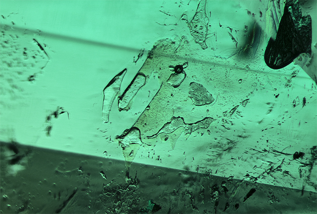
Figure 1 offers a great example of this. This inclusion scene in emerald shows an irregular cavity. When illuminated with a long-wave UV flashlight (figure 2), it is immediately evident that the cavity is filled with a substance that displays a chalky blue fluorescence. Observation under long-wave UV light also makes it easier to observe gas bubbles in the filled areas.
.jpg)


