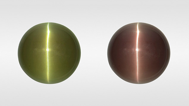
Cat’s-Eye Alexandrite with Unique Inclusion Pattern

Chatoyancy is an optical effect caused by light reflecting off dense concentrations of parallel needles or hollow tubes in cabochon-cut gem materials. To maximize chatoyancy, the base of the cabochon should be cut parallel to the inclusions to produce an even effect across the dome. In some cases, the parallel inclusions are not distributed throughout the stone and are only in zones or narrow bands. In such cases the cutter must orient the needles with care to produce a cat’s-eye.A chatoyant cabochon, weighing 21.22 ct and measuring 17.75 × 17.50 × 6.77 mm, was recently submitted to GIA’s Tokyo laboratory for identification service. This stone displayed a change of color (figure 1) from brownish green in fluorescent light to brownish purple in incandescent light. Standard gemological testing resulted in a spot refractive index (RI) reading of 1.75, a specific gravity (SG) of 3.74, and strong trichroism. In addition to these properties, heterogeneous needle-like inclusions and infrared and Raman spectroscopy revealed that the stone was natural chrysoberyl.
What is notable about this stone is that all the needle-like inclusions that cause the chatoyancy are confined to a narrow layer at the very bottom of the cabochon (figure 2). The rest of the stone is nearly free of inclusions. The cutter obviously knew to orient what must have been a narrow layer of needles at the bottom of the cabochon so that the dome would form a complete cat’s-eye. Some synthetic or imitation stones have an engraved foil backing or an engraved base to produce a cat’s-eye or star in much the same way. Raman spectroscopic analysis indicated that the needle-like inclusions were rutile (figure 3). While the boundary between the included and inclusion-free areas looks sharp, the stone is not assembled.
.jpg)


