Resin-Coated and Clarity-Enhanced Aquamarine Pendant
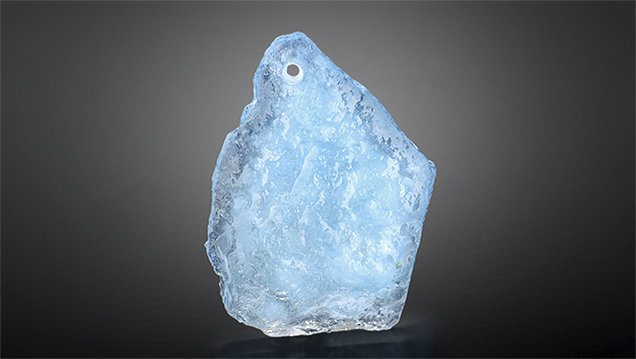
The Carlsbad laboratory received a 34.24 ct light greenish blue free-form pendant (figure 1) measuring 33.40 × 25.75 × 6.40 mm. One side of the pendant was flat and polished, and the other side had a rough surface. Standard gemological testing gave a refractive index (RI) of 1.570–1.580. The pendant had the same reaction to long-wave and short-wave UV light: blue fluorescence in the center and green fluorescence around the edges of the stone. Examination with a standard gemological microscope revealed a blue and orange flash effect most clearly visible from the flat side (figure 2, left), which is diagnostic of clarity enhancement. Ultraviolet/visible/near-infrared (UV-Vis-NIR) spectra were consistent with natural aquamarine, with absorption peaks at 370 and 425 nm and an absorption band at ~830 nm (figure 2, right). Raman spectroscopy showed the presence of polymer on the rough side of the stone and aquamarine on the polished flat side. Although emeralds are the variety of beryl most commonly associated with clarity enhancement, filled aquamarine has previously been reported in the Chinese market (J. Li et al., “Polymer-filled aquamarine,” Fall 2009 G&G, pp. 197–199).


Only one side, the rough side, was coated with polymer resin. Viewed in the microscope, this side appeared to have a thin transparent layer on the surface. Trapped air bubbles were visible with magnification (figure 3, left), and areas with the coating had a higher luster than the polished flat side. Fourier-transform infrared (FTIR) spectroscopy taken from the rough side revealed strong diagnostic peaks at ~3100–2850 cm–1 (figure 3, right).
Although clarity enhancement and coating of beryl gemstones are not uncommon, this material is rarely seen in a gemological laboratory. Both treatments are readily identified, but careful examination of every stone is important to identify them.
.jpg "YMAL GIA Ed")


