Separating Glass-Filled Rubies Using the DiamondView

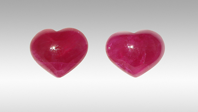
The treatment of gemstones using various filler materials is not new. In the case of corundum, silica glass was often observed filling rubies from approximately 1980 to the early 2000s, when it was largely replaced by high-lead-content glass in early 2004 (S.F. McClure et al., “Identification and durability of lead glass–filled rubies,” Spring 2006 G&G, pp. 22–34) which has since become the mainstream artificial filler in corundum. The Lai Tai-An Gem Lab in Taipei recently examined a pair of rubies (figure 1) that were quickly determined to be glass filled with the assistance of DiamondView imaging. In our experience, the DiamondView detects the presence of glass, although it does not distinguish between different types of glasses. This note shows how the DiamondView can be used to observe the presence and, perhaps more importantly, the degree of filling in rubies.
The two heart-shaped cabochons weighed 6.81 and 6.89 ct and measured approximately 10.78 × 13.55 × 5.39 mm and 11.36 × 13.10 × 5.47 mm, respectively. Each semitransparent stone exhibited a purplish red color and fluoresced a strong red to long-wave ultraviolet radiation while exhibiting a weak red reaction to short-wave ultraviolet irradiation. Standard gemological testing showed spot refractive indices of 1.76, and specific gravities of 3.94 and 3.93, respectively, which were lower than regular values and raised suspicion that the stones were filled, while advanced analysis by energy-dispersive X-ray fluorescence (EDXRF) and Fourier-transform infrared (FTIR) spectroscopy and Raman microscopy revealed data consistent with ruby. Magnification with a gemological microscope revealed natural fluid and fingerprint inclusions, but some evidence of a filler was also observed in the fractures.


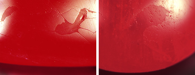
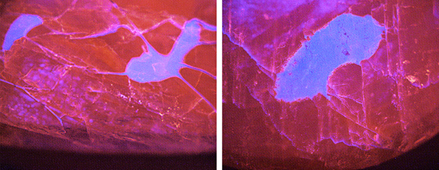
Under the DiamondView’s visible light settings, some surface-reaching filler features and filled “cavities” were also revealed. Notable differences in the surface reflectance between the filling material and surrounding corundum (figure 2) were clearly evident. After exposure to the DiamondView’s short-wave UV radiation, the treated nature of both rubies was even more apparent as the reaction not only showed the obvious cavities but the extent of the filler (in blue) running throughout the stones (figure 3). The 6.81 ct ruby revealed an even greater degree of filling than its twin, especially on the base where a greater concentration of filling material was noted.
As the DiamondView results prove, filler materials penetrate or remain on the fractures of the treated material (ruby in this case) under certain treatment conditions. Hence, some filling treatments may be revealed more clearly when exposed to the ultra-short-wave energy of the DiamondView. Chemical analysis of a filled area using EDXRF detected Si and Ba as the major elements. Minute levels of Pb were also obtained. The results proved the filler was a glass composed of these elements, which were not detected when other filler-free areas were analyzed.
Artificial glass is frequently used as a filling material in gemstones. The variable chemical compositions of these different glasses help improve the appearance (lower visibility of fractures) of the host after treatment. While the vast majority of glasses and other fillers, both organic and inorganic, can be detected with a loupe or microscope, the DiamondView is a useful identification aid should the need arise.
.jpg)


