Cape Diamond with Yellow Phosphorescence

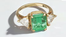
Phosphorescence in a cape diamond is very rare, especially after exposure to long-wave UV light. The New York laboratory recently examined a cape diamond exhibiting yellow phosphorescence after exposure to a long-wave UV source. The 0.75 ct Faint yellow-green round brilliant is shown in figure 1.
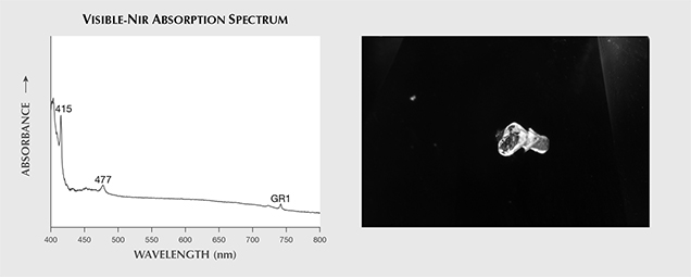
Cape absorption peaks were observed at 415 and 477 nm, along with a GR1 peak (figure 2, left). The IR spectrum confirmed that this was a type Ia diamond, with nitrogen aggregates detected in the one-phonon region. Hydrogen-related peaks were also detected at 1405, 2785, 3107, 3237, 4169, and 4496 cm–1. Brown radiation stains were found on the girdle. The diamond contained contacted partially graphitized omphacite crystals with cuboctahedral morphology (figure 2, right). Strong blue fluorescence and medium yellow phosphorescence were observed under a desktop long-wave UV light source. Under short-wave UV, medium yellow fluorescence was noted but phosphorescence was absent. DiamondView imaging (<225 nm excitation) also showed greenish blue fluorescence but no phosphorescence. Fiber-optic illumination revealed the blue “transmission” luminescence that occurs when a strong light travels through a diamond. The UV-Vis spectrum of such diamonds shows an absorption peak at 415 nm and a luminescence peak at the lower energy end of the peak. Such is the case with this stone.

Nearly 30 years ago, a bicolor diamond with a near-colorless central portion and light yellow tips was reported in G&G (Winter 1989 Lab Notes, p. 237). It showed very weak chalky yellow phosphorescence for approximately 10 seconds after the long-wave UV lamp was turned off. This yellow phosphorescence was observed throughout the stone regardless of its bicolor nature. Its central colorless region exhibited weak cape lines and strong blue transmission, similar to the green diamond in this note. Unlike our sample, the bicolor diamond also showed very weak yellow phosphorescence to short-wave UV. A chameleon diamond may display strong yellow phosphorescence to a long-wave UV light source (Summer 1992 Lab Notes, p. 124; Spring 2000 Lab Notes, pp. 60–61). Our sample was not a chameleon diamond, however.
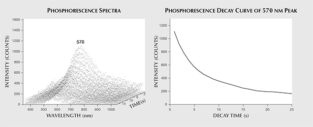
Greenish yellow phosphorescence in a chameleon diamond has been systematically measured using a spectrometer (see S. Eaton-Magaña et al., “Fluorescence spectra of colored diamonds using a rapid, mobile spectrometer,” Winter 2007 G&G, pp. 332–351). The peak maximum recorded for the chameleon diamond was 557 nm. We used an Ocean Optics USB2000 charge-coupled device (CCD) spectrometer similar to the one described in Eaton-Magaña et al. (2007), but with a different UV source. In place of a deuterium source, we used GIA’s new LED desktop long-wave UV (365 nm excitation) light source. A broad peak at approximately 570 nm, which is responsible for the yellow phosphorescence, was observed in the emission spectra (figure 3, left). The phosphorescence decay curve at 570 nm, representing the rate of decreasing intensity with time, is plotted in figure 3 (right). Half-life is defined as the time required for the initial peak intensity to decrease to one-half its original value (again, see Eaton-Magaña et al., 2007). The half-life value measured for our sample was 5.0 seconds.

Yellow phosphorescence is a very rare feature in cape diamonds. The defect responsible for this optical feature remains unknown.


