Assembled Cultured Blister Pearl with an Unusual Component

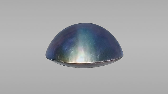
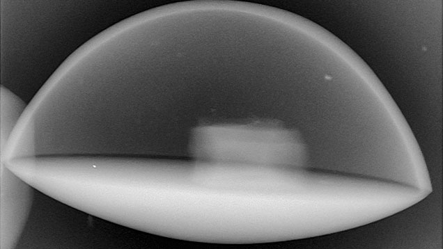
GIA’s Bangkok laboratory recently examined a blue-violet assembled cultured blister pearl (mabe) weighing 2.44 ct and measuring 10.23 × 5.86 mm (figure 1). The specimen had the appearance of a typical mabe pearl, exhibiting a boundary line between the nacre face and shell base. 2D microradiography showed a component with an unusual fluted shape, with radio-translucency similar to that of the CaCO3 nacre and the shell (figure 2).

GIA has examined the characteristics of mabe pearls used in commercial jewelry over the years (see Lab Notes: Summer 1981, pp. 104–105; Summer 1991, pp. 111–112; Fall 1991, p. 177; Summer 1992, pp. 126–127; Fall 1992, pp. 195–196; Fall 1996, p. 210), but none of these notes mentioned this unusual component. In Bangkok, this feature has been seen five times in mabe pearls submitted for testing since 2010; questions about its identity and purpose have been raised since the first observation.
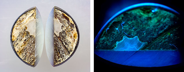
Out of curiosity, one client decided to cut their sample in half in order to examine the interior in more detail (figure 3, left). The two halves consisted of a thin nacre dome top, which was coated with a dark layer on the inner surface; an artificial resinous material; and the white fluted object bordering the shell base. The fluted object was translucent, and a clear spiderweb structure was visible at 10× magnification. When exposed to standard short-wave UV radiation (254 nm) and examined in the DiamondView, this object showed a moderate white blue reaction that followed the spiderweb pattern (figure 3, right).

Further investigation with Raman spectroscopy using a 488 nm laser revealed a calcite spectrum with peaks at 159, 285, 716, 1088, and 1750 cm–1, similar to white coral (Corallium secundum). A small peak related to carotenoid pigments was present at 1020 cm–1 (e.g., J. Urmos et al., “Characterization of some biogenic carbonates with Raman spectroscopy,” American Mineralogist, Vol. 76, 1991, pp. 641–646).
Based on the spiderweb structure, radio-translucency, calcite spectrum, and general appearance, the object appeared to be a biogenic carbonate. White coral was the first material considered. Further research revealed accounts of sea urchin sections used in some assembled cultured blister pearls from Bali, Indonesia, in “Kuta” pearls (E. Strack, Pearls, Ruhle-Diebener-Verlag, Stuttgart, Germany, 2006, p. 605). Regardless of identity, the reason for including this material within some mabe pearls remains uncertain. It does not appear related to either the formation of the mabe pearl component or its weight and stability.


