Micro-World Gems & Gemology, Winter 2022, Vol. 58, No. 4

Garnet with Apatite Inclusions

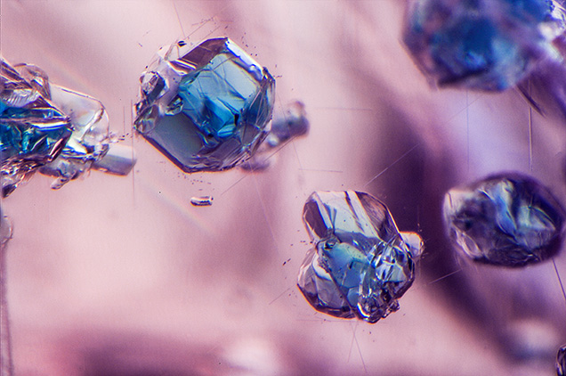
Figure 1. Left: This 3.07 ct purplish pink “dragon” garnet, as seen in daylight-equivalent lighting, displays eye-visible blue apatite inclusions. Right: Red fluorescence in the garnet due to chromium, as seen using long-wave UV. Photos by Jessa Rizzo; courtesy of Ravenstein Gem Co.
Figure 2. Blue apatite inclusions and rutile needles were prominent throughout the “dragon” garnet reportedly from a new find in Africa. Photomicrograph by Nathan Renfro; field of view 2.40 mm.

The authors recently examined a 3.07 ct garnet sample acquired from Ravenstein Gem Co. by author NR. This gem material, marketed online as “dragon” garnet as an allusion to the mythical creature’s changing eye color, was reportedly from a new find at an undisclosed locality in Africa. It is also notable that Lotus Gemology has reported similar material from Tanzania (Summer 2022 G&G Micro-World, pp. 226–227). The garnet was a delicate purplish pink color under daylight-equivalent lighting (figure 1, left) and showed a fairly strong red fluorescence when exposed to long-wave ultraviolet light (figure 1, right). Also of note, this sample, as well as many of the other examples showcased online, contained vibrant blue inclusions of apatite (figure 2) as well as typical needle-like silk and minute fluid inclusions.

Figure 3. The garnet’s UV-Vis-NIR spectrum suggests the color results mainly from manganese, iron, chromium, and vanadium. Narrow bands at 409, 422, and 430 nm are related to manganese in the garnet structure, while absorption bands related to iron are at ~465 and 488 nm (Z. Sun et al., “Quantitative definition of strength of chromophores in gemstones and the impact on color change in pyralspite garnets,” <i>Color Research and Application</i>, Vol. 47, 2022, pp. 1134–1154). A broad band centered at ~572 nm is from vanadium and chromium absorption.
Figure 3. The garnet’s UV-Vis-NIR spectrum suggests the color results mainly from manganese, iron, chromium, and vanadium. Narrow bands at 409, 422, and 430 nm are related to manganese in the garnet structure, while absorption bands related to iron are at ~465 and 488 nm (Z. Sun et al., “Quantitative definition of strength of chromophores in gemstones and the impact on color change in pyralspite garnets,” Color Research and Application, Vol. 47, 2022, pp. 1134–1154). A broad band centered at ~572 nm is from vanadium and chromium absorption.
Figure 4. The photoluminescence spectrum collected from the garnet using a 514 nm laser revealed chromium-related emission consistent with the observed red fluorescence under long-wave UV excitation.

Gemological testing revealed a refractive index measurement of 1.741 and a hydrostatic specific gravity of 3.81. Further gemological testing with laser ablation–inductively coupled plasma–mass spectrometry revealed the major composition to be pyrope (61.5 mol.%), spessartine (29.0 mol.%), grossular (6.5 mol.%), and almandine (2.6 mol.%), a chemistry consistent with pyralspite-series garnets. Notable trace elements in significant quantities were chromium (660 ppmw) and vanadium (343 ppmw) in addition to the rare earth elements yttrium (1206 ppmw), erbium (222 ppmw), and ytterbium (420 ppmw). The ultraviolet/visible/near-infrared (UV-Vis-NIR) spectrum revealed absorption bands that were consistent with the chemical analysis (figure 3), indicating that the color of the garnet results from a mixture of manganese, iron, chromium, and vanadium. Raman analysis confirmed the blue inclusions as apatite. It is also notable that the garnet sample showed a weak color change in daylight compared to various non-standardized, commercially available LED types of lighting, changing from purplish pink to pink-orange. Photoluminescence testing with a 514 nm laser revealed a strong chromium-related emission, consistent with the red fluorescence observed with long-wave UV exposure (figure 4).

The strong red fluorescence, color change under certain LED lights, and presence of significant rare earth elements and vibrant blue apatite inclusions make it quite interesting for any gem collector.

Jessa Rizzo is a senior staff gemologist, Nathan Renfro is senior manager of colored stone identification, and Ziyin Sun is a senior research associate, at GIA in Carlsbad, California.