Quartzite Bangle with Fuchsite Inclusions
Jadeite jade bangles are always popular in the Chinese market. With high prices and rising demand for these products, a bangle with an appearance similar to jadeite was encountered recently.
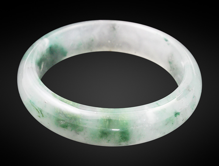
The bangle was submitted to the National Gold-Silver Gem & Jewelry Quality Inspection Center (Sichuan) for identification. It was semitransparent with a white and green bodycolor resembling that of jadeite (figure 1). However, standard gemological testing showed a refractive index of 1.54 and a specific gravity of 2.67, values that were both significantly below those of jadeite and more in line with quartz. It showed no reaction under Chelsea filter. Using a polariscope, the material was found to be composed of polycrystalline aggregates. Microscopic examination revealed a granular aggregate structure and different microcrystalline mineral inclusions.

The identity was further confirmed by analytical testing. Fourier-transform infrared (FTIR) reflectance spectroscopy showed peaks at 489, 537, 691, 778, 798, 1105, and 1178 cm–1 that were consistent with the characteristic spectrum of quartzite (sometimes referred to as “quartzose”) (figure 2) (X. Jin et al., “Gemmological and vibrational spectrum characteristics of micaceous quartzose jade ‘strawberry crystal’,” Journal of Gems & Gemmology, Vol. 23, No. 3, 2021, pp. 20–28). Energy-dispersive X-ray fluorescence (EDXRF) analysis revealed trace amounts of iron throughout and chromium exclusive to the green inclusions.
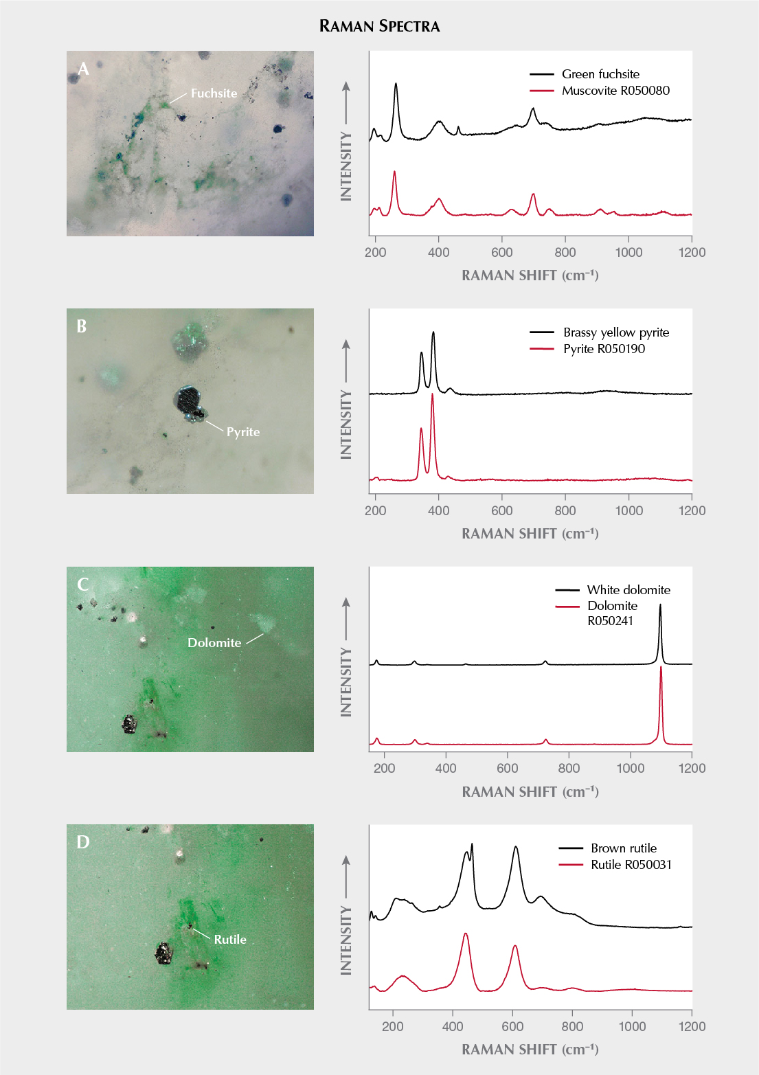
These green flaky inclusions were slightly oriented. Raman combined with EDXRF analysis identified them as fuchsite (K(Al,Cr)2(AlSi3O10)(OH)2), a green chromium-colored variety of muscovite (figure 3A) (Fall 2013 GNI, pp. 183–184). Additionally, brassy yellow inclusions in a range of sizes were confirmed as pyrite (figure 3B) using Raman spectroscopy. Raman also identified some white blocky inclusions in a nearby area as dolomite (figure 3C), as well as some tiny brown rutile inclusions found as irregular, randomly scattered grains (figure 3D).

Ultraviolet/visible spectroscopy corresponding to the green areas showed two broad absorption bands at about 423 and 620 nm, with a weak peak at 684 nm (figure 4). These features have been assigned to Cr3+ in fuchsite.
To our knowledge, fuchsite contained in quartzite is usually uniformly scattered in the form of tiny flakes. The uneven distribution of green fuchsite that resulted in patches of green in this bangle is noteworthy. The patchy green spots resemble the color concentrations often observed in jadeite. Despite the bangle’s resemblance to jadeite, gemological tests such as refractive index, microscopic observation, and infrared and Raman spectroscopy readily identify it.


