Color-Change Spessartine Garnet: A First Report

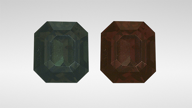
GIA’s Carlsbad laboratory recently examined a 2.11 ct faceted stone (figure 1) that showed strong color change from dark green under daylight-equivalent lighting to dark red under incandescent illumination. Standard gemological testing revealed a refractive index that was over the 1.80 RI limit of the GIA desktop refractometer. This was the first example of a color-change garnet with an over-the-limit RI reading examined by GIA. Fluorescence was inert to long-wave and short-wave UV light. The stone did not show any pleochroism with the dichroscope. Using a handheld spectroscope, we observed a cutoff at about 460 nm and a broad band centered at about 575 nm. Microscopic examination revealed only a small fracture and strong internal strain. High-order interference colors were observed in cross-polarized light from the strain along grain boundaries.
Laser ablation–inductively coupled plasma–mass spectrometry (LA-ICP-MS) results showed that the stone was composed predominantly of spessartine (80.37 mol.%), with minor contributions from other end members of the garnet species (see supplementary table 1). It is classified as a spessartine garnet based on the criteria in the pyrope-spessartine-almandine ternary diagram (figure 2, left) modified from Stockton and Manson (“A proposed new classification for gem-quality garnets,” Winter 1985 G&G, pp. 205–218). The RI and SG could also be calculated based on chemical composition (W.A. Deer et al., Rock-Forming Minerals: Orthosilicates, Vol. 1A, Geological Society of London, 1982, pp. 485–488). The calculated RI and SG—plotted in figure 2, right—were 1.797 and 4.182, respectively. The calculated RI value was consistent with the measured RI value. In 2001, Krzemnicki et al. reported a garnet composed of 76.9 mol.% spessartine and 4.2 mol.% goldmanite with a color change from orange-yellow under daylight-equivalent lighting to reddish orange under incandescent illumination based on the colorimetric data in the article, although color change from brownish green to brownish was observed visually (“Colour-change garnets from Madagascar: Comparison of colorimetric with chemical data,” Journal of Gemmology, 2001, Vol. 27, No. 7, pp. 395–408). The high spessartine and goldmanite composition of the stone is similar to our stone reported here, but with much weaker color change.


To understand why this spessartine garnet showed a color-change phenomenon, we collected an ultraviolet/visible/near-infrared (UV-Vis-NIR) spectrum (the visible portion of the spectrum is shown in figure 3), which was then used to quantitatively calculate the stone’s colorimetric coordinates (L*, a*, and b*; see table 2 at www.gia.edu/gems-gemology/summer-2019-labnotes-color-change-spessartine-garnet-icp-table2.pdf) under different lighting conditions. The strong absorption below 450 nm is caused by Mn2+ (R.K. Moore and W.B. White, “Electronic spectra of transition metal ions in silicate garnets,” Canadian Mineralogist, Vol. 11, No. 4, 1972, pp. 791–811). A wide absorption band centered at about 585 nm is caused mainly by V3+ (C.A. Geiger et al., “Single-crystal IR- and UV/VIS-spectroscopic measurements on transition-metal-bearing pyrope: the incorporation of hydroxide in garnet,” European Journal of Mineralogy, Vol. 12, No. 2, 2000, pp. 259–271), which produced two transmission windows: one in the blue-green part of the spectrum (figure 3, window A) and the other in the red (figure 3, window B). This was the cause of the color change in this spessartine garnet, as incandescent illumination highlighted the red transmission window and daylight-equivalent lighting highlighted the blue-green window. Color pairs under CIE D65 illumination (representing daylight-equivalent lighting) and CIE A illumination (representing incandescent illumination) were calculated and are shown in figure 3, above transmission window A and B. The darker color pair (top row), directly calculated from the measured UV-Vis-NIR spectrum, closely matched the dark appearance of the stone (again, see figure 1) with L*(lightness) around 14.5. It was the strong absorption generated by very high Mn and V concentration, along with the stone’s relatively large size, that produced a long light path length. To better observe the saturation and hue of the color pair, the authors changed the L*to 50 and reproduced the color pair with the same a*and b*values (bottom row). The hue difference of the color pair is easier to see with increased lightness.


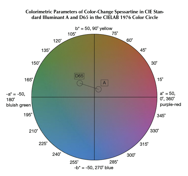
One way to judge the quality of a color-change stone is to plot the color pair in the CIELAB 1976 color circle. Good color-change pairings show a large hue angle difference, small chroma difference, and large chroma values (Z. Sun et al., “How to facet gem-quality chrysoberyl: Clues from the relationship between color and pleochroism, with spectroscopic analysis and colorimetric parameters,” American Mineralogist, Vol. 102, No. 8, 2017, pp. 1747–1758). The color coordinates L*, a*, and b*(again, see table 2) of the stone under CIE D65 illumination and CIE A illumination were plotted in the CIELAB 1976 color circle in figure 4. The spessartine showed a low saturation under CIE A illumination.

Color change has been reported previously in pyrope-spessartine (K. Schmetzer et al., “Color-change garnets from Madagascar: Variation of chemical, spectroscopic and colorimetric properties,” Journal of Gemmology, Vol. 31, No. 5-8, 2009, pp. 235–282), in pyrope (Z. Sun et al., “Vanadium- and chromium-bearing pink pyrope garnet: characterization and quantitative colorimetric analysis,” Winter 2015 G&G, pp. 348–369), and in grossular (Spring 2018 GNI, pp. 233–236). To the authors’ knowledge, this was the first color-change spessartine reported.


