Fossil Insect in Opal
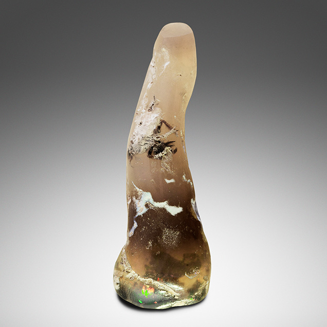
The authors recently examined a most unusual opal. The mottled brown polished free-form stone appeared to contain a trapped insect, which one might expect to find in fossil amber or copal (figure 1). However, noticeable play-of-color made it obvious that the host material for the insect was precious opal, which was further confirmed by standard gemological testing. Its refractive index of 1.45, weak white long-wave fluorescence, and Raman spectrum were all consistent with natural opal. No microscopic evidence of any type of treatment was detected.

Play-of-color was strongest in the darker brown portions toward the base but also appeared in shallow fissures around the insect’s appendages. The insect broke the surface, resulting in some of its legs and underside being cut through during polishing (figure 2, left). An apparent set of mouth parts was clearly observed, but the position of some pits on the surface partially obscured the view. Fine hairs, or setae, were found along the legs (figure 2, center). The setae closest to the surface were surrounded by a grainy white material that resembled desiccated opal. A slightly contorted abdomen was observed alongside a pair of rear legs (figure 2, right).
Precious opal formation is not fully understood, but several mechanisms have been proposed (B.Y. Lynne et al., “Diagenesis of 1900-year-old siliceous sinter (opal-A to quartz) at Opal Mound, Roosevelt Hot Springs, Utah, U.S.A.,” Sedimentary Geology, Vol. 179, Nos. 3-4, 2005, pp. 249–278; B. Pewkliang et al., “The formation of precious opal: Clues from the opalization of bone,” The Canadian Mineralogist, Vol. 46, No. 1, 2008, pp. 139–149). One possible explanation is that low-pH groundwater percolated through the soil, accruing colloidal silica into a silica-rich fluid. This gel then underwent polymerization within cavities and voids to form microspheres of opal-A. One could imagine a scenario where the insect was entrapped by an intrusion of this gel, rapidly enveloping the insect and allowing it to avoid decomposition.
While fossilized organic matter in the form of roots and twigs has been found in precious opal—most notably in Virgin Valley, Nevada, and Wollo, Ethiopia—this is the first confirmed case of an opal-encased insect that the authors have examined. We were not able to find any other recorded findings, although there have been rumors of them. It is our hope that more specimens are discovered so we can more thoroughly study these exceptional rarities.


