Pearls Marketed as “Venezuelan Pearls”
The term “Venezuelan pearls” is applied to natural saltwater pearls harvested from the Pinctada imbricata mollusk, also known as the Atlantic pearl oyster. They are found primarily in the western Atlantic, from Bermuda and Florida to northern South America. The mollusk size ranges from 5 to 7 cm and produces pearls ranging from less than 2 mm (seed pearls) up to 9 mm (CIBJO Pearl Guide, 2022). Some traders claim that many pearls sold as “Venezuelan pearls” in the world markets today, mainly India and the Middle East, are sometimes mixed with pearls from other Pinctada species (typically lower- to medium-quality Pinctada maxima pearls). Others claim that actual pearls from Venezuela are also sold as “Basra pearls,” which are sourced from Pinctada radiata and are highly valued in India and the Middle East due to their appearance and luster.
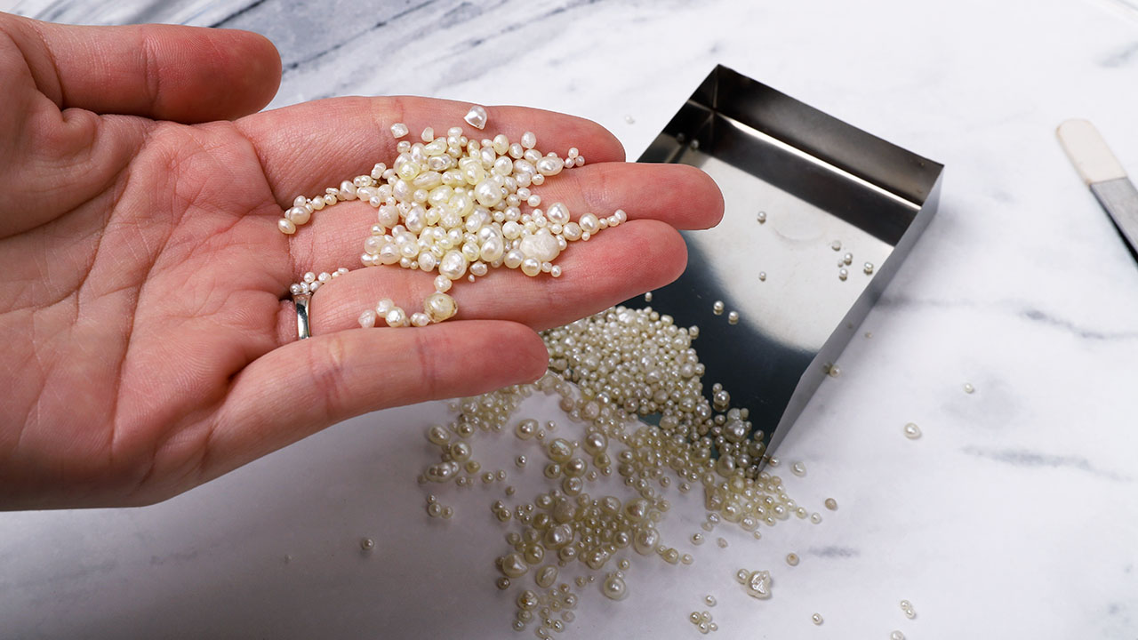
GIA’s Mumbai laboratory studied a parcel of 988 reportedly “Venezuelan pearls” from one of the main suppliers of this material (figure 1). The internal structures were examined with real-time X-ray microradiography (RTX), and 97 samples were selected for further study. The majority of the button-shaped and semi-baroque pearls were white to light cream and cream in color, and most exhibited strong orient. Their weight ranged from 0.05 to 1.06 ct, with sizes from 2.05 × 1.88 mm to 6.87 × 5.23 × 4.23 mm. Most of the samples exhibited a good luster with a smooth surface, and a few revealed minor surface blemishes. Under high magnification, the samples showed various patterns of overlapping aragonite platelets on the surface, which are typically observed in nacreous pearls.
Energy-dispersive X-ray fluorescence on the 97 selected samples revealed low manganese levels ranging from below detection limit to 29 ppm, with a few ranging from 32.5 to 69.8 ppm. Higher strontium levels were also observed, from 1177 to 2988 ppm, with a few samples ranging from 3056 to 3437 ppm. When exposed to X-ray fluorescence, most of the samples showed an inert reaction, while a few exhibited an extremely faint yellowish green reaction. Under long-wave ultraviolet radiation, all the pearl samples luminesced various degrees of bluish green.
Raman spectroscopy revealed a doublet at 701/704 cm–1 and a peak at 1086 cm–1, with a few pearls showing a weak peak at 1464 cm–1 indicative of aragonite. Additionally, the spectra of one light cream and one cream pearl showed weak polyenic pigment peaks at 1135 and 1530 cm–1. These observations were previously documented for pearls from Pinctada fucata (akoya) and Pinctada radiata (A. Al-Alawi et al., “Saltwater cultured pearls from Pinctada radiata in Abu Dhabi (United Arab Emirates),” Journal of Gemmology, Vol. 37, No. 2, 2020, pp. 164–179). Due to the pearls’ smaller size, ultraviolet/visible/near-infrared spectra were collected for only 54 of them. Some of these displayed weak bands around 320, 395, and 420 nm, while others showed a prominent feature at 460 nm, similar to previously studied samples from Pinctada imbricata.
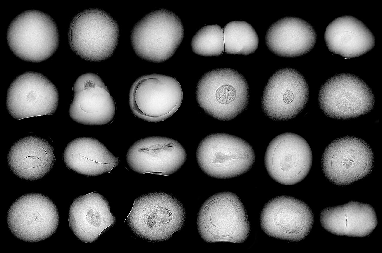
RTX analyses revealed a combination of structures that were typical of those observed in natural pearls from Pinctada species, as shown in figure 2. In this study, around 44% of the pearls revealed faint gray dense cores surrounded by growth arcs and organic-rich gaps at their outer edges. The samples displayed tight and minimal internal growth structures, some with thicker concentric growth arc formations following the shape of the pearl, representing approximately 16% of the group. Additionally, around 13% showed multi-nuclei structures, exhibiting a combination of different types of cores, while a few revealed interesting crystalline structures.
The most challenging to identify were void structures and linear features, which were found in only 6% of the samples. Some of these were long features that, to some extent, mirrored the outline of the pearl. The linear features originated from the tip of the pearls and were surrounded by a few growth arcs. These were similar to the odd linear structures observed in natural pearls from various mollusk species in GIA’s research database. A total of 18% of the pearls revealed irregular centrally positioned voids of varying sizes. Voids are frequently observed in saltwater non-bead cultured (NBC) pearls, particularly those from Pinctada species. Therefore, it was important to further study these samples using X-ray computed microtomography (μ-CT) imaging, which indicated that many voids were filled with fine, light gray organic-rich matter, unlike the empty voids typically observed in NBC pearls (A. Homkrajae et al., “Internal structures of known Pinctada maxima pearls: Cultured pearls from operated marine mollusks,” Fall 2021 G&G, pp. 186–205).
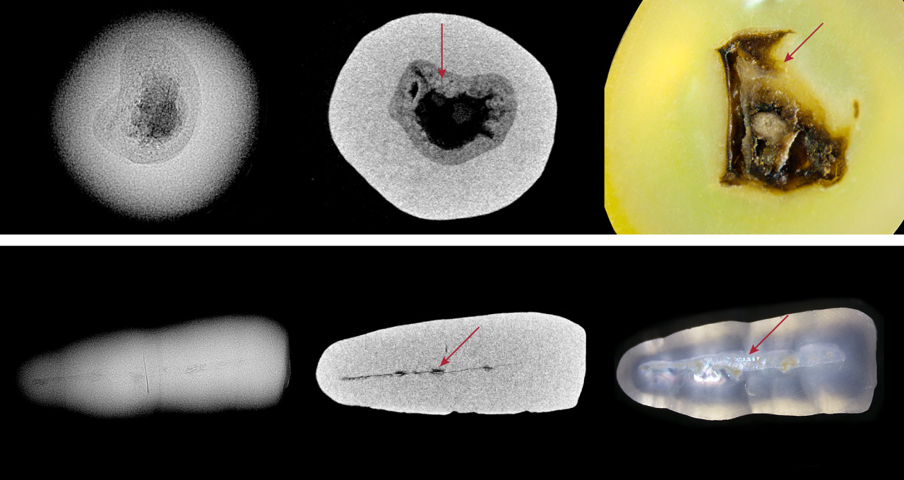
To further study these challenging internal structures, two pearls were ground down. The dark gray void-like area seen in the RTX image of pearl A was composed of a solid organic material that appeared as a light and dark brown area in the cross section (figure 3, top row). Pearl B, which showed an elongated linear structure in the RTX image, had a hollow space with organic-rich pockets (figure 3, bottom row).
Based on comparison with research samples from GIA’s database, some of the samples from this lot had internal structures similar to those from Pinctada imbricata, but others displayed overlap with structures found in Pinctada radiata and Pinctada maxima. Although a few samples showed borderline void and linear internal structures, similar to NBC pearls from Pinctada maxima, the majority had structures seen in natural pearls from the other Pinctada species. These findings suggest that the potential mixing of these pearls, whether intentional or accidental, may be attributed to their remarkably similar external appearance. Consequently, more advanced testing utilizing laser ablation–inductively coupled plasma–mass spectrometry will be necessary to effectively differentiate between the pearls from the different Pinctada species mollusks mixed within the studied lot of pearls. Therefore, GIA’s ongoing research serves as a valuable resource for identifying pearls from different mollusks.


