A Minute Foraminifera Shell in a Natural Non-Nacreous Black Pearl

Nacre can form around nearly any irritant that infiltrates a shell, resulting at times in pearls with unique internal structures. In very rare cases, foraminifera shell (also known as “test”) can serve as the nuclei of pearls produced by different mollusks (Spring 2023 GNI, pp. 143–145). These tiny single-celled marine microorganisms can be found on the seafloor and are usually about half a millimeter to one millimeter long.
GIA’s Mumbai laboratory recently received a lot of non-nacreous brown and black pearls of various shapes, collected by a Bahraini diver over a period of six years. The lot was submitted for scientific examination and contained pearls from both the Pinctada radiata and Pinna species (pen pearls) fished from Bahrain. Real-time X-ray microradiography (RTX) revealed different types of cores and acicular (radial) structures in most of the pearls. One very intriguing structure was observed in a black near-round pearl weighing 0.02 ct and measuring 1.55 × 1.43 mm, reportedly from a Pinctada radiata mollusk (figure 1).
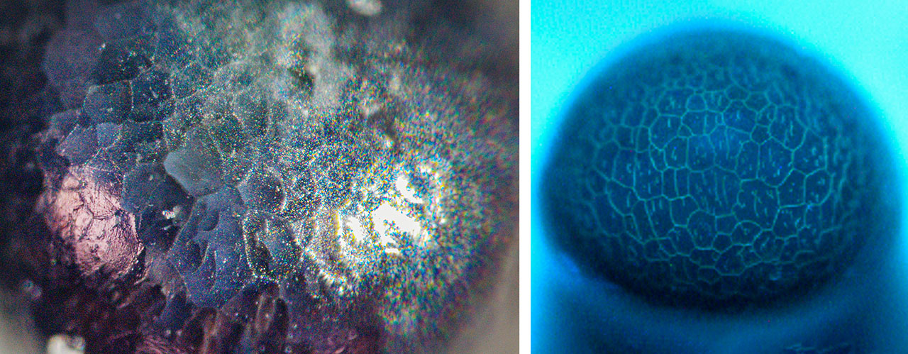
The pearl had a glossy appearance with a few pits and no surface-reaching cracks. Viewed under high magnification with a fiber-optic light, it showed a non-nacreous surface, presenting cellular outlines with fine small lines within each cell (figure 2, left). When exposed to X-ray fluorescence, the pearl showed an inert reaction. Energy-dispersive X-ray fluorescence spectrometry revealed a manganese level of 76 ppm and a higher strontium level of 1160 ppm, indicative of a saltwater origin. Under long-wave and short-wave ultraviolet radiation, the pearl showed an inert reaction. However, DiamondView imaging revealed a remarkable cellular structure within. A bluish reaction was observed at the boundaries outlining the cellular columnar structures with thin, short lines within the cells (figure 2, right). This cellular surface structure appeared different from the pseudohexagonal cellular outline found in pen pearls (N. Sturman et al., “Observations on pearls reportedly from the Pinnidae family (pen pearls),” Fall 2014 G&G, pp. 202–215). Based on the authors’ previous observations, this type of structure is more typically found on the surface of non-nacreous pearls from the Pinctada radiata mollusk.
Raman spectroscopy using 514 nm laser excitation was performed on two spots. The first spot revealed peaks at 150 and 280 cm−1 with the associated band at 1086 cm−1. The second spot also revealed a peak at 1086 cm−1 associated with aragonite and calcite, but there were no other clear peaks to identify which of the two calcium carbonate polymorphs was present in that area of the pearl.
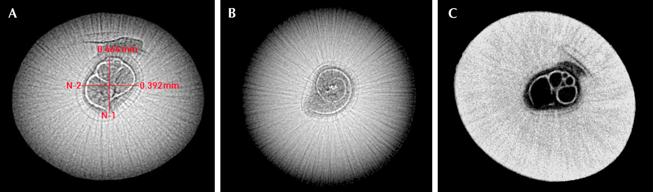
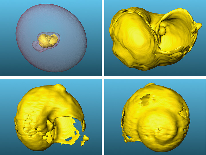
Interestingly, RTX imaging and X-ray computed microtomography (μ-CT) analysis revealed a minute foraminifera shell at the pearl’s core, measuring approximately 0.44 × 0.38 mm. The shell exhibited a double thin-walled structure, surrounded by an organic-rich area and an acicular (radial) structure with fine growth arcs crossing it. The radiating acicular structure covered the entire area of the pearl (figure 3, A and C). When observed from the top view, the RTX image showed the spiral shape of the shell’s apex (figure 3B). Given its chamber shape and tiny size, the shell was identified as a foraminifera (K. Kimoto et al., “Precise bulk density measurement of planktonic foraminiferal test by X-ray microcomputed tomography,” Frontiers in Earth Science, Vol. 11, 2023, article no. 1184671). Additionally, μ-CT scan images were rendered using specialized software (C. Zhou et al., “New 3-D software expands GIA’s pearl identification capabilities,” GIA Research News, May 13, 2016), which showed a clearer visualization of the external morphology of the foraminifera shell within the pearl (figure 4), as shown in the video below.
Black non-nacreous pearls from Pinctada radiata are extremely rare, and it is very uncommon to see such intriguing internal structures with foraminifera shells surrounded by acicular structures. The presence of this non-nacreous seed pearl with such a structure adds to GIA’s comprehensive research database, contributing to the ongoing studies of unique formations found in natural pearls from the wild.


