Research Article Search Results
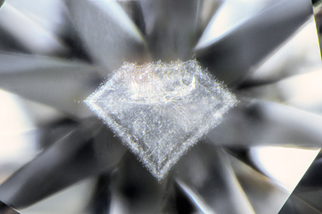
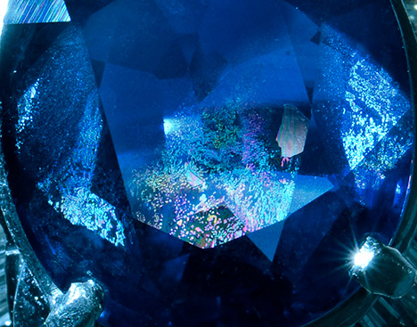
Diamond-Shaped Cloud in Diamond
In a delightful juxtaposition, a diamond-shaped cloud inclusion appears in the center of a 0.53 ct round brilliant diamond.
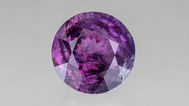
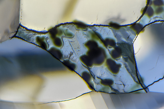
Rare Faceted Hexagonite
The Carlsbad laboratory recently examined an unusually large faceted hexagonite with iridescent cleavage cracks and needle-like structures.
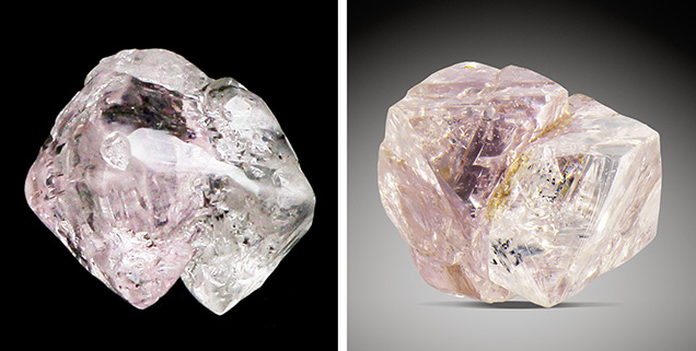
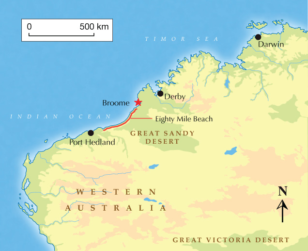
Bicolor Rough Diamond Crystals
Two Australian rough diamonds demonstrate both pink and colorless sections in the same crystal.
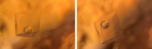
Opal with Fluid Inclusion and Mobile Bubble
The Carlsbad laboratory recently examined a Mexican opal containing a rare fluid inclusion with a freely moving gas bubble.
Expanded Diamond Surface Due to Radiation Staining
The author examined a 0.56 ct Fancy grayish bluish green diamond with prominent green radiation stains.
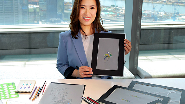
Fourth Annual Gianmaria Buccellati Foundation Award
A graduate of GIA’s Hong Kong Jewelry Design program hopes her award-winning design will help raise environmental awareness.
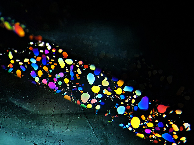
Thin-Film Fluid Inclusions in Aquamarine
Oblique fiber-optic illumination of thin-film fluid inclusions in Brazilian aquamarine reveals a kaleidoscopic play of interference colors.
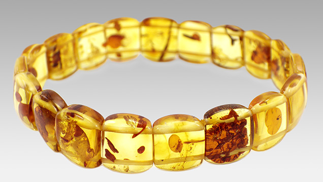
Unusual Fluorescence of a Color-Enhanced Amber Bracelet
Abnormal fluorescence supports the likelihood that this amber bracelet had been “baked” to enhance its color.
Virtual Report on the Industry in 2021
An overview of how the gem and jewelry trade, from miners to manufacturers, and from designers to dealers, dealt with the many challenges posed by the pandemic.
Remarkably Large Iridescent Healed Fissures Resembling Play-of-Color in Sapphire
Attractive iridescence creates a beautiful “play-of-color” effect on dendritic healed fissures in a blue sapphire.
Somewhere in the Rainbow Finds a Home at the Alfie Norville Gem and Mineral Museum
A beautiful new gem and mineral museum in Tucson is home to the stunning Somewhere in the Rainbow collection.
Internal Structures of Known Pinctada maxima Pearls: Natural Pearls from Wild Marine Mollusks
Presents the internal characteristics of natural P. maxima pearls, obtained from real-time microradiography (RTX) and X-ray computed microtomography (µ-CT) analysis of 774 samples.


