
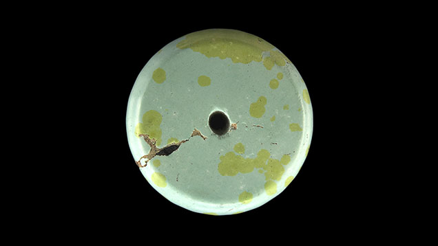
Recently, author YYL encountered a unique specimen of turquoise characterized by yellow raindrop-like patterns in the Zhushan County turquoise market in Hubei Province, China. The sample exhibited a blue-green substrate, with bright yellow spots distributed individually and in clusters across its surface. Both the substrate and the spots were opaque, with the individual spots nearly round and measuring between 2 mm and 1 cm in size (figure 1).
Fourier-transform infrared (FTIR) analysis of both the substrate and the raindrop-like spots revealed turquoise vibrational peaks. Two absorption peaks attributed to a stretching vibration were noted at 2858 and 2979 cm–1, indicative of wax (Fall 2015 GNI, pp. 343–345). The stronger absorptions for these two features in the substrate suggested a higher wax content, likely from residual wax after polishing due to the sample’s lower hardness.
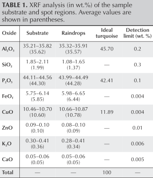
To determine the compositions of the substrate and the spots, four points in each of these regions were tested with X-ray fluorescence (XRF) spectroscopy using a quantitative method developed by the School of Jewellery laboratory, China University of Geosciences, Wuhan, with the total content of major oxides ranging from 98 to 100 wt.%. The results indicated that the primary chemical compositions of the yellow spots and the substrate were largely consistent (table 1). Specifically, the average Al2O3 content was 35.57 wt.% for the yellow spots and 35.62 wt.% for the substrate. Similarly for the yellow spots and substrate, the CuO content was 10.78 wt.% and 10.60 wt.%, respectively, while the total iron oxide (FeOT) content was 6.44 wt.% and 5.85 wt.%, respectively.
Trace element analysis was performed using laser ablation–inductively coupled plasma–mass spectrometry on three spots each in the substrate and raindrop areas across both sides of the sample, with the results demonstrating broadly consistent element enrichment patterns between the substrate and raindrops. No significant concentration differences between the yellow raindrop regions and the blue-green substrate were observed for the trace elements analyzed, with the exception of vanadium and uranium, which were both significantly higher in concentration in the yellow regions.
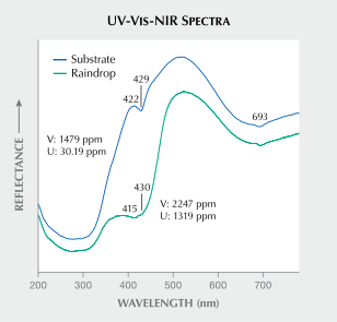
The ultraviolet/visible/near-infrared (UV-Vis-NIR) reflection spectra of the substrate exhibited characteristics typical of turquoise (figure 2). In contrast, the yellow spots displayed a strong absorption band at 415 nm, accompanied by a shoulder band at 430 nm. The violet-blue region showed significant absorption, while the red region exhibited a broader absorption band, albeit with lower intensity compared to the blue-violet region. From a compositional perspective, the primary distinction between the substrate and the yellow spots lies in their uranium and vanadium contents. Uranium exhibits a strong absorption band at 415 nm in the violet region, forming a broad absorption feature. The low-energy end of this absorption extends to approximately 460 nm, which is attributed to the magnetic dipole transition of the uranyl ion (A.N. Platonov and C. Jin, “Color of uranyl-containing minerals,” World Nuclear Geoscience Journal, Vol. 1, 1975, pp. 38–42). An increase in vanadium content causes the absorption related to iron ions (Fe3+) at 422 nm to shift toward the red region, resulting in an absorption feature at 430 nm (X. Wang and Y. Guo, “The impact of trace metal cations and absorbed water on colour transition of turquoise,” Royal Society Open Science, Vol. 8, No. 2, 2021, article no. 201110). In summary, absorption in the violet region is primarily caused by uranium, vanadium, and iron, while copper contributes to absorption in the red region. These elemental contributions collectively account for the intense yellow coloration observed in the spots.
Although this raindrop turquoise exhibits distinct color variations, there were no significant differences observed in the FTIR spectral characteristics or bulk chemical composition between the substrate and the yellow spots. However, when looking at trace element chemistry, significantly higher levels of vanadium and uranium were identified in the spots compared to the substrate. A slightly higher concentration of iron was also observed in the yellow spots.
Yunyu Liu and Quanli Chen are affiliated with the Gemmological Institute, China University of Geosciences, Wuhan.
Recently, author YYL encountered a unique specimen of turquoise characterized by yellow raindrop-like patterns in the Zhushan County turquoise market in Hubei Province, China. The sample exhibited a blue-green substrate, with bright yellow spots distributed individually and in clusters across its surface. Both the substrate and the spots were opaque, with the individual spots nearly round and measuring between 2 mm and 1 cm in size (figure 1).
Fourier-transform infrared (FTIR) analysis of both the substrate and the raindrop-like spots revealed turquoise vibrational peaks. Two absorption peaks attributed to a stretching vibration were noted at 2858 and 2979 cm–1, indicative of wax (Fall 2015 GNI, pp. 343–345). The stronger absorptions for these two features in the substrate suggested a higher wax content, likely from residual wax after polishing due to the sample’s lower hardness.
To determine the compositions of the substrate and the spots, four points in each of these regions were tested with X-ray fluorescence (XRF) spectroscopy using a quantitative method developed by the School of Jewellery laboratory, China University of Geosciences, Wuhan, with the total content of major oxides ranging from 98 to 100 wt.%. The results indicated that the primary chemical compositions of the yellow spots and the substrate were largely consistent (table 1). Specifically, the average Al2O3 content was 35.57 wt.% for the yellow spots and 35.62 wt.% for the substrate. Similarly for the yellow spots and substrate, the CuO content was 10.78 wt.% and 10.60 wt.%, respectively, while the total iron oxide (FeOT) content was 6.44 wt.% and 5.85 wt.%, respectively.
Trace element analysis was performed using laser ablation–inductively coupled plasma–mass spectrometry on three spots each in the substrate and raindrop areas across both sides of the sample, with the results demonstrating broadly consistent element enrichment patterns between the substrate and raindrops. No significant concentration differences between the yellow raindrop regions and the blue-green substrate were observed for the trace elements analyzed, with the exception of vanadium and uranium, which were both significantly higher in concentration in the yellow regions.
The ultraviolet/visible/near-infrared (UV-Vis-NIR) reflection spectra of the substrate exhibited characteristics typical of turquoise (figure 2). In contrast, the yellow spots displayed a strong absorption band at 415 nm, accompanied by a shoulder band at 430 nm. The violet-blue region showed significant absorption, while the red region exhibited a broader absorption band, albeit with lower intensity compared to the blue-violet region. From a compositional perspective, the primary distinction between the substrate and the yellow spots lies in their uranium and vanadium contents. Uranium exhibits a strong absorption band at 415 nm in the violet region, forming a broad absorption feature. The low-energy end of this absorption extends to approximately 460 nm, which is attributed to the magnetic dipole transition of the uranyl ion (A.N. Platonov and C. Jin, “Color of uranyl-containing minerals,” World Nuclear Geoscience Journal, Vol. 1, 1975, pp. 38–42). An increase in vanadium content causes the absorption related to iron ions (Fe3+) at 422 nm to shift toward the red region, resulting in an absorption feature at 430 nm (X. Wang and Y. Guo, “The impact of trace metal cations and absorbed water on colour transition of turquoise,” Royal Society Open Science, Vol. 8, No. 2, 2021, article no. 201110). In summary, absorption in the violet region is primarily caused by uranium, vanadium, and iron, while copper contributes to absorption in the red region. These elemental contributions collectively account for the intense yellow coloration observed in the spots.
Although this raindrop turquoise exhibits distinct color variations, there were no significant differences observed in the FTIR spectral characteristics or bulk chemical composition between the substrate and the yellow spots. However, when looking at trace element chemistry, significantly higher levels of vanadium and uranium were identified in the spots compared to the substrate. A slightly higher concentration of iron was also observed in the yellow spots.
Yunyu Liu and Quanli Chen are affiliated with the Gemmological Institute, China University of Geosciences, Wuhan.