
ABSTRACT
This study investigated the gemological characteristics of red and orange spinel from Mogok in Myanmar and from Lang Chap and An Phu in Vietnam. In terms of standard gemological properties, all samples displayed a refractive index ranging from 1.710 to 1.728, specific gravity of 3.528 to 3.692, and red fluorescence under ultraviolet light. Advanced gemological methods, including ultraviolet/visible/near-infrared spectroscopy, electron probe microanalysis, and laser ablation–inductively coupled plasma–mass spectrometry, were employed to investigate chemical compositions, particularly focusing on chromophores and determination of diagnostic origin criteria. Both red and orange spinel samples exhibited chemical properties close to stoichiometric spinel. It was found that the diverse coloration in red and orange samples resulted from variations in chromium-to-vanadium ratios. The studied red spinels were chromium-rich, with chromium-to-vanadium ratios ≥1.7, whereas the orange spinels were vanadium-rich, with chromium-to-vanadium ratios ≤1.0. Trace element plots of titanium versus zinc for red spinel revealed separation between the three sources (Mogok, Lang Chap, and An Phu). Regarding orange spinel, plotting of vanadium versus zinc showed separation for Lang Chap and An Phu samples but due to the wide range of composition found in the Mogok samples, there was overlap between samples from Mogok and the two Vietnamese sources. Therefore, separation of the orange spinel from these sources is more difficult and will require investigating additional samples.
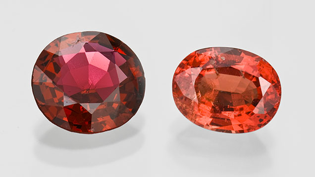
Spinel, as a gemstone, has continued to increase in popularity in recent years (figure 1). Despite high demand for this gem among collectors and in the jewelry trade, obtaining fine-quality spinel has proven challenging due to limited availability. Consequently, the price of spinel has consistently trended upward; moreover, the gemstone’s geographic origin also plays a pivotal role in pricing. The origin of spinel is becoming an increasingly significant determinant of its value and is expected to soon be as significant as for emerald, ruby, and sapphire. For instance, blue spinel from Tanzania and Vietnam is more renowned than that from Madagascar or Myanmar, while red spinel from Myanmar and Vietnam is more highly regarded than that from other origins. However, reliable origin determination of spinel requires dependable data. Therefore, a comprehensive analysis of the physical and chemical characteristics of spinel from various origins is necessary. Color variation alone is insufficient to determine country of origin; similar appearing pink spinel might come from quite different geological environments. For instance, Tajikistan spinel occurs in forsterite lenses within magnesian skarn (Liu et al., 2022), whereas the spinels studied here from the main commercial deposits in Myanmar (Mogok) and Vietnam (Lang Chap and An Phu) are both situated in marble hosts. These marbles formed under high-grade (granulite facies) metamorphic conditions and tectonic activity linked to the Himalayan orogeny (Garnier et al., 2005; Khoi et al., 2011; Huong et al., 2012; Fallick et al., 2019; Phyo et al., 2019; Chankhantha et al., 2020). The similarities in the marble hosts may result in comparable trace element chemistries and identical mineral inclusion assemblages. Consequently, these complicate the separation and origin determination of the studied spinels.
Spinel is the magnesium- and aluminum-containing member (MgAl2O4) of the spinel group of minerals with the general formula (X2+)(Y3+)2O4 (Sickafus et al., 1999) and crystallizes in the isometric (cubic) system. For spinel, the refractive index and specific gravity are approximately 1.72 and 3.55, respectively (Deer et al., 2013). However, solid solutions may form where different metals substitute for magnesium and aluminum in the X2+ and Y3+ sites and may slightly change those physical properties, introducing color. For instance, red, pink, and orange spinel may be attributed to the presence of Cr3+ and V3+ presumably in the octahedral (Y3+) site, whereas blue spinel is attributed to Co2+ and/or Fe2+ predominantly in the tetrahedral (X2+) site. However, spinel with a blue hue originating from cobalt tends to exhibit a more saturated and brighter color compared to spinel with a blue hue from iron. Chauviré et al. (2015) and Krzemnicki et al. (2023) also reported the presence of cobalt in blue spinel from Vietnam and Tanzania. Purple spinel can form when combinations of chromium and vanadium are accompanied by iron and/or cobalt substituting in the X2+ and/or Y3+ sites (Peretti and Günther, 2003; Chauviré et al., 2015; Andreozzi et al., 2019; Belley and Palke, 2021; Krzemnicki et al., 2023). However, the flexible nature of the octahedral and tetrahedral structures in spinel allows both sites to potentially accommodate either 2+ or 3+ ions (D’Ippolito et al., 2015; Andreozzi et al., 2019; Zhang et al., 2023). Investigations using quantitative analytical methods for trace element concentrations have been employed to separate natural from synthetic spinel, as well as to identify treatments (Saeseaw et al., 2009; Pluthametwisute et al., 2022). However, the trace element profiles of spinel from various origins remain largely unexplored.
GEOLOGICAL BACKGROUND
Mogok, Myanmar. Mogok is a significant global source of ruby, sapphire, and spinel. Gem mining has taken place in both primary and secondary deposits in this region since the fifteenth century (Iyer, 1953). Mogok is located in central Myanmar and is geologically part of the Mogok Metamorphic Belt (MMB) (figure 2) (Iyer, 1953; Phyo et al., 2019; Chankhantha et al., 2020), which experienced two major metamorphic events. The first occurred during the late Cretaceous to Paleocene, while the second took place during the late Eocene to Oligocene. Both events are linked to the collision between the Indian and Asian tectonic plates, which resulted in the formation of the Himalayan mountain range (Iyer, 1953; Searle et al., 2007, 2020; Phyo et al., 2019). The geologic setting of the Mogok region is highly complex, mainly consisting of high-grade metamorphic schists, gneisses, marble, calc-silicate rocks, and quartzite. Additionally, various igneous intrusions are present, including gem crystal–containing pegmatites and ultramafic rocks (Iyer, 1953; Thu, 2007; Phyo et al., 2019). Spinel has typically been found in marbles associated with metamorphism in granulite facies. Other minerals discovered in these marble formations include apatite, chondrodite, clinohumite, diopside, dravite, forsterite, graphite, phlogopite, pyrite, and corundum (ruby) (Zaw et al., 2015; Chankhantha et al., 2020). Moreover, anhydrite, apatite, calcite, chondrodite, dolomite, graphite, ilmenite, magnesite, forsterite, phlogopite, potassium feldspar, pyrite, quartz, rutile, sulfur, titanite, uraninite, and zircon have also been found as mineral inclusions in Mogok spinel (Phyo et al., 2019). Although mineral inclusions can provide valuable information for gemological characterization and origin determination, most of the samples examined in this study were free of inclusions, which limited detailed inclusion analysis.
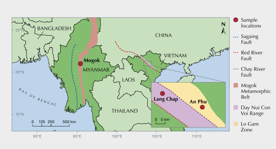
Lang Chap, Vietnam. Here, gem spinel has been mined mostly from secondary deposits, beginning in February 2010. This area is particularly renowned for red to orange spinel (Khoi et al., 2011; Huong et al., 2012). The small village of Lang Chap is located in the Luc Yen District, Yen Bai Province, within the Day Nui Con Voi range. This area is positioned between the Red River Fault to the southwest and the Chay River Fault to the northeast (Trinh et al., 2012; again, see figure 2). In the Red River Fault zone, two main tectonic events contributed to the formation of gemstones; the Indosinian orogeny resulted from the collision between the Yangtze craton and the Indochinese shield in the Upper Proterozoic (Kušnír, 2000); the subsequent Himalayan orogeny produced similar high-grade metamorphic conditions to those of the MMB. Additionally, spinel from Lang Chap is associated with high-grade metamorphic rocks under amphibolite facies’ conditions, including calc-silicates, gneisses, and marbles (Malsy et al., 2012; Chauviré et al., 2015; Giuliani et al., 2017). As a newly discovered deposit, detailed information on mineral inclusions in Lang Chap spinel is limited.
An Phu, Vietnam. This renowned gem mining location is also in Yen Bai Province and is known for high-quality spinel. These spinels formed within marble during granulite facies metamorphism; other minerals found in these marbles include chlorite, clinohumite, forsterite, pargasite, and phlogopite (Garnier et al., 2005; Fallick et al., 2019). In addition, inclusions of apatite, calcite, dolomite, goethite, graphite, magnesite, potassium feldspar, titanite, and zircon were discovered in these spinels (Giuliani et al., 2003). An Phu is part of a geological terrane, the Lo Gam zone, to the east of the Red River Fault (Malsy et al., 2012; again, see figure 2). Two significant tectonic processes in the Lo Gam zone were crucial to the formation of gemstones along the Red River Fault area. These were the Indosinian orogeny, resulting from the collision between the Yangtze craton and the Indochinese shield during the Upper Proterozoic, and the subsequent Himalayan orogeny, which is also associated with the metamorphic evolution of the MMB (Garnier et al., 2005; Chauviré et al., 2015; Giuliani et al., 2017; Chankhantha et al., 2020).
MATERIALS AND METHODS
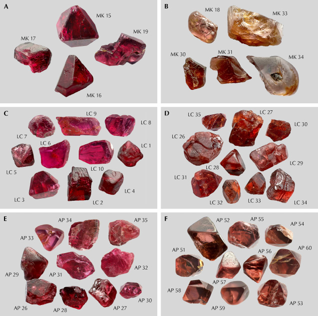
Samples. All spinel samples for this study were acquired by the authors from either miners or local markets around the mining areas. Author DS bought all Mogok spinels directly from the local market around the mining area, and author DTAV bought spinels directly from the local miners in Lang Chap and An Phu, Vietnam. Red and orange spinels were meticulously selected and categorized based on their color and hue according to the Munsell color system. The red spinels’ color codes ranged from 2.5R to 5R, and the orange spinels’ color codes ranged from 7.5R to 10R. The red spinel collection comprised 13 samples from Mogok, 25 samples from Lang Chap, and 25 samples from An Phu. The orange spinel collection included 14 samples from Mogok, 25 samples from Lang Chap, and 25 samples from An Phu. All samples were labeled using abbreviations of locations followed by a number (e.g., MK 17). Double-sided polished wafers were fabricated from all 127 spinel samples for standard gemological testing, including determination of refractive index (RI), specific gravity (SG), ultraviolet (UV) reaction, and optical character with a polariscope. Furthermore, ultraviolet/visible/near-infrared (UV-Vis-NIR) spectroscopy, electron probe microanalysis (EPMA), and laser ablation–inductively coupled plasma–mass spectrometry (LA-ICP-MS) were applied to select samples representing each unique color (figure 3).
UV-Vis-NIR Spectroscopy. Absorption spectra in the UV-Vis-NIR range (250–1500 nm) were collected by a Perkin-Elmer Lambda 1050 spectrophotometer at the Gem and Jewelry Institute of Thailand (GIT). Scan speed was set at 405 nm/min with a resolution of 3 nm. The thin double-sided polished wafer samples (≤4 mm thick) allowed for the use of transmission mode. The absorption coefficient was calculated using the formula α = 2.303A/t, where α represents the Napierian true absorption coefficient in cm–1, A is the decadic true absorbance (unitless), and t is the thickness of the sample in centimeters (Dubinsky et al., 2020).
EPMA. A JEOL model JXA-8100 electron probe microanalyzer at the Department of Geology, Chulalongkorn University, was used to determine major and minor oxide compositions. Standard materials for calibration included synthetic quartz (SiO2) for silicon, synthetic rutile (TiO2) for titanium, synthetic corundum (Al2O3) for aluminum, lead vanadium germanium oxide (PbV Geoxide) for vanadium, synthetic eskolaite (Cr2O3) for chromium, gadolinium gallium garnet (GGG) for gallium, fayalite (Fe2SiO4) for iron, synthetic periclase (MgO) for magnesium, synthetic manganosite (MnO) for manganese, cobalt oxide (CoO) for cobalt, zinc oxide (ZnO) for zinc, and wollastonite (CaSiO3) for calcium. An acceleration voltage of 15 kV and a probe current of about 2.5 × 10⁻⁸ A with a focused electron beam (<1 μm) were applied to all analytical spots. Detection limits were empirically below 0.005 wt.%. Three spots were analyzed for each sample.
LA-ICP-MS. Trace element analyses were performed with LA-ICP-MS. This unit was comprised of a Thermo Scientific iCAP RQ ICP-MS attached to an Elemental Scientific Laser NWR-213 (laser ablation unit) based at GIT. The parameters employed for ICP-MS were a carrier gas flow of 0.75 L/min of helium, a plasma gas flow of 14 L/min of argon with plasma power set at 1550 W, and a radiofrequency generator supply of 40 V. Supplementary parameters for laser ablation included an output energy of 0.246 mJ, fluence around 10 J/cm2, laser energy set at 65%, spot size of 85 μm, repetition rate of 10 Hz, dwell time of 60 s, and three analyzed spots for each sample. External standard calibration was performed using NIST SRM 610 (with lithium, beryllium, and boron concentrations of 485.0, 465.9–466.1, and 357.0 ppm, respectively) and NIST SRM 612 (with lithium, beryllium, and boron concentrations of 41.5, 36.2–38.4, and 35.0 ppm, respectively). The following masses were collected: 49Ti, 51V, 53Cr, 55Mn, 56Fe, 59Co, 60Ni, 67Zn, and 69Ga. All analyses were reported as ppmw and converted to ppma on the basis of the molecular weight per atom approach, which is as follows: Molecular weight of spinel (MgAl2O4) is divided by the total number of atoms per formula unit (7 atoms) to obtain the average mass per atom. This value is then divided by the atomic weight of the target element and multiplied by its ppmw value to obtain the atomic concentration (ppma).
RESULTS
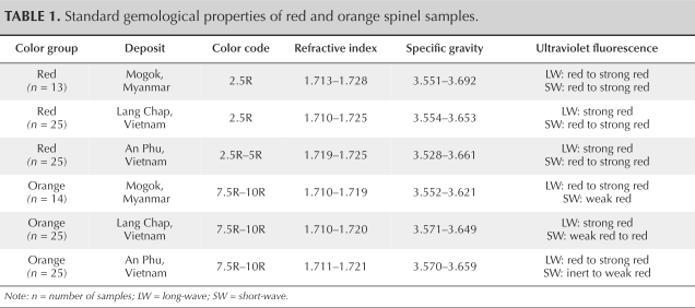
Standard Gemological Properties. A summary of the standard gemological properties for the red and orange spinels tested is presented in table 1. The samples ranged in RI from 1.710 to 1.728, all displaying single refraction. The SG values varied within a range of 3.528 to 3.692. Notably, all red spinel samples displayed red fluorescence under both long-wave (365 nm) and short-wave (254 nm) UV, while some orange spinel samples exhibited red fluorescence under long-wave UV but were inert or exhibited weak to medium red fluorescence under short-wave UV.
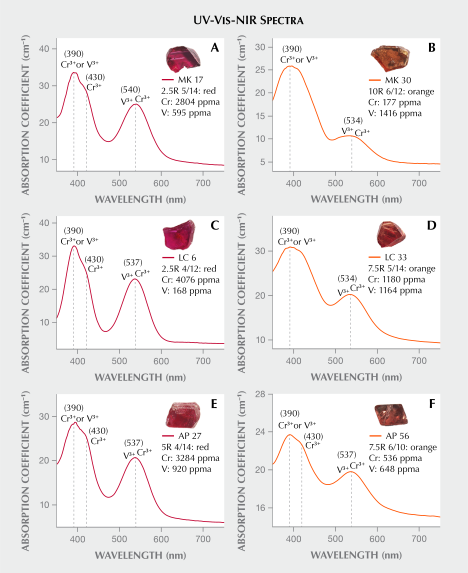
UV-Vis-NIR Spectroscopy. UV-Vis-NIR absorption spectra corrected for path length of all red and orange spinel samples showed absorption peaks at 390 nm and 534–540 nm (figure 4), attributed to overlapping contributions from Cr3+ and V3+. Notably, the absorption feature associated with Cr3+ in red spinel shows a shoulder at 430 nm. In addition, orange spinels exhibited a reduced absorption intensity from 470 to 600 nm compared to their red counterparts, while also displaying a less pronounced peak at 534–540 nm. Moreover, their Cr3+ absorption range (350–490 nm) was broader than that observed in red spinels (350–470 nm). The referenced absorption peaks of Cr3+ and V3+ are from Belley and Palke (2021) and D’Ippolito et al. (2015).
Distinct absorption bands were evident for red spinel in the violet range (350–470 nm) and green to yellow range (500–600 nm), resulting in transmission in the blue and red regions of the spectrum (figure 4, A, C, and E). On the other hand, orange spinels exhibited similar absorption in the violet range (350–470 nm), extending into part of the blue region (490 nm), along with reduced absorption in the green to yellow range (500–600 nm), resulting in increased transmission in the orange region, as depicted in figure 4, B, D, and F.
The representative red spinel sample (LC 6 in figure 4C) revealed a chromium-rich and vanadium-poor composition, with 4076 ppma chromium and 168 ppma vanadium, and exhibited a peak at 390 nm, a shoulder peak at 430 nm, and a distinct absorption intensity at 534–540 nm. In contrast, the representative orange spinel sample (MK 30 in figure 4B) yielded chromium-poor and vanadium-rich contents of 177 ppma chromium and 1416 ppma vanadium and showed a peak at 390 nm, a broader peak width compared to the red spinel, and a much reduced peak at 534–540 nm.
Mineral Chemistry. EPMA. Representative EPMA analyses and the resulting calculated stoichiometric formulas based on four oxygen atoms are presented in tables 2 and 3. Moreover, Fe2+ and Fe3+ were recalculated using the method described by Droop (1987). According to the classification of Haggerty (1991) and Deer et al. (2013), spinels with major elements of magnesium and aluminum in an approximate stoichiometric ratio of 1:2 are classified as spinel (MgAl2O4).
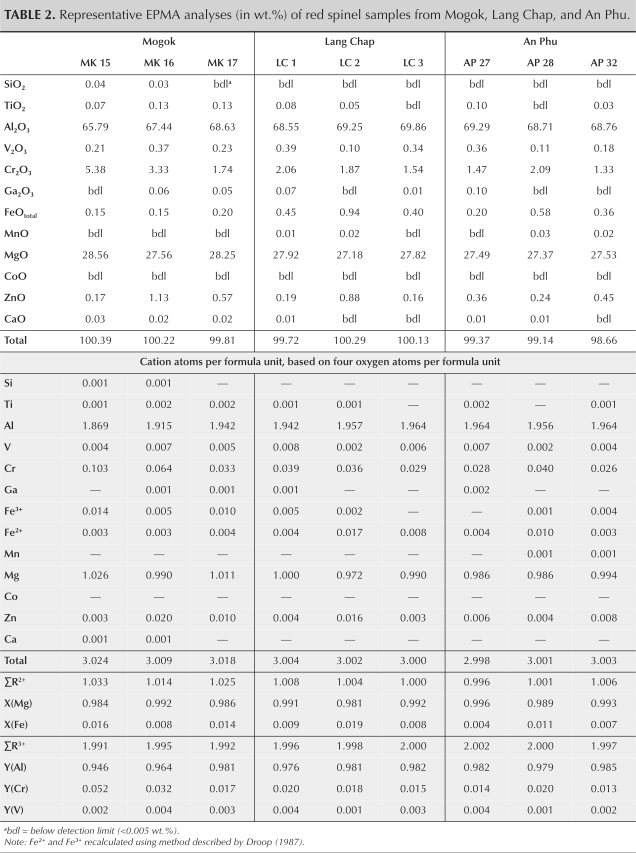
Chemical analyses of red spinel samples from Mogok, Lang Chap, and An Phu demonstrated that all contained Al2O3 (65.79–69.86 wt.%) and MgO (27.18–28.65 wt.%). Consequently, the red samples exhibited 1.869–1.964 Al3+ atom per formula unit (apfu), incompletely occupying the octahedral site (2 apfu). The presence of Cr3+ (0.026–0.103 apfu) was observed, substituting for Al3+ in the octahedral site. Consequently, the mole fraction at the tetrahedral site showed that XMg2+ ranged from 0.981 to 0.996, while XFe2+ ranged from 0.004 to 0.019. At the octahedral site, YAl3+ ranged from 0.946 to 0.985, YCr3+ ranged from 0.013 to 0.052, and YV3+ ranged from 0.001 to 0.004 (table 2).
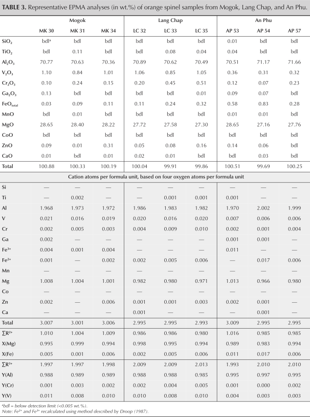
Orange spinel samples from Mogok, Lang Chap, and An Phu were predominantly comprised of Al2O3 (70.36–71.66 wt.%) and MgO (27.16–28.65 wt.%). Therefore, recalculated Al3+ ions (1.968–2.002 apfu) occupied most of the octahedral sites. In addition, V3+ (0.006–0.021 apfu) was observed, substituting for Al3+ in the octahedral site. As a result, the mole fraction at the tetrahedral site showed that XMg2+ ranged from 0.983 to 0.993, while XFe2+ ranged from 0.001 to 0.017. At the octahedral site, YAl3+ ranged from 0.985 to 0.997, YCr3+ ranged from 0 to 0.005, and YV3+ ranged from 0.003 to 0.011 (table 3).
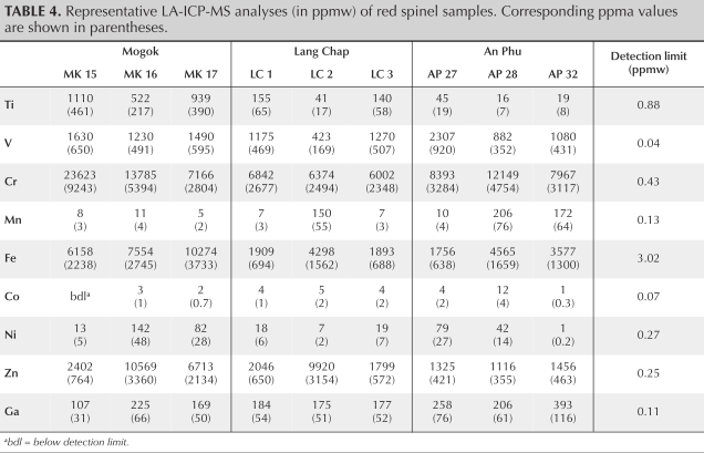
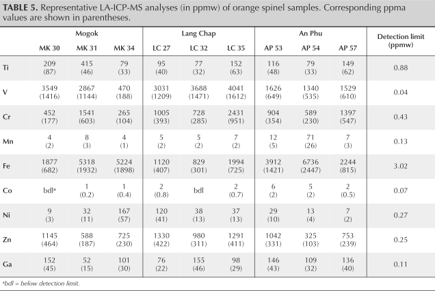
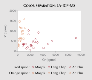
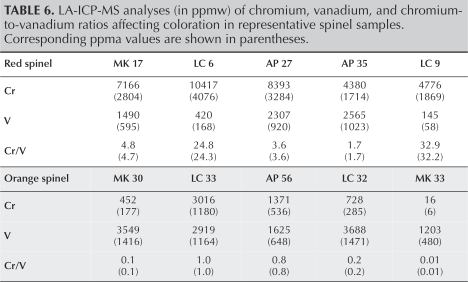
LA-ICP-MS. Representative trace analyses of impurities in red and orange spinel samples are presented in tables 4 and 5, respectively. It is well recognized that the red and orange colorations in spinel are primarily attributed to chromium and vanadium, as seen in these samples. The relationship between chromium and vanadium is illustrated in figure 5, highlighting the significantly higher chromium contents in red spinel compared to vanadium, while the reverse is observed in orange spinel. Notably, the chromium-to-vanadium ratios are ≥1.7 for red spinel and ≤1.0 for orange spinel (table 6). These ratios were determined using both ppmw and ppma and yielded similar values because chromium and vanadium have very similar atomic weights.
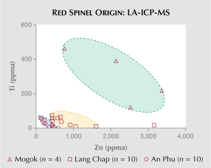
Trace analyses of red spinel (table 4) determined a vanadium range of 169 to 920 ppma, a chromium range of 2348 to 9243 ppma, and an iron range of 638 to 3733 ppma. The chromium-to-vanadium ratios displayed a range of 1.7 to 32, indicating chromium is always predominant over vanadium (mostly ≥1.7 times). Additionally, titanium versus zinc plots distinctly highlight a variation among different sources, with a decreasing trend in titanium content from Mogok to Lang Chap and An Phu, respectively (figure 6).
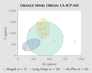
The orange spinel samples (table 5) were determined to have a range of vanadium from 188 to 1612 ppma, chromium from 104 to 951 ppma, and iron from 301 to 2447 ppma. In particular, chromium-to-vanadium ratios were clearly ≤1.0 (table 6). Iron content in orange spinel appears to be lower than in red spinel. Additionally, plots of vanadium versus zinc are distinctively different between Lang Chap and An Phu orange spinel; however, they both overlap with Mogok spinel (figure 7).
DISCUSSION
The colors observed in spinel are predominantly influenced by trace elements substituting in tetrahedral and/or octahedral sites, resulting in spin-allowed or spin-forbidden transitions that contribute to a diverse range of colors (Chauviré et al., 2015; Andreozzi et al., 2019). Specifically, the red and orange hues in spinel are attributed to Cr3+ and V3+ in spin-allowed transitions, leading to the absorption of light within the violet spectrum (350–470 nm) and the green to yellow spectrum (500–600 nm). The red spinel samples, characterized by chromium-to-vanadium ratios ≥1.7, exhibited a distinct peak at 390 nm (with a shoulder peak at 430 nm) and a band at 534–540 nm, resulting in a pronounced red hue. Conversely, the studied orange spinel, with chromium-to-vanadium ratios ≤1.0, exhibited a distinct peak at 390 nm (without a 430 nm shoulder peak) and a band at 534–540 nm, showed enhanced light absorption in the blue to green spectrum (350–490 nm), and displayed decreased absorption intensity in the green to yellow spectrum (500–600 nm), producing a distinct orange color due to the partial cancellation of the red range by the complementary green range. For example, samples LC 6, MK 17, and AP 27, with chromium-to-vanadium ratios of 24, 4.7, and 3.6, respectively (table 6), clearly demonstrated this effect. In contrast, sample MK 30, with a color code of 10R 6/12 and a chromium-to-vanadium ratio of 0.01, displayed a more prominent orange color compared to samples LC 33 and AP 56, which have chromium-to-vanadium ratios of 1.0 and 0.83, along with color codes of 7.5R 5/14 and 7.5R 6/10, respectively (again, see figure 4, D and F). Notably, spinel samples with chromium-to-vanadium ratios between 1.0 and 1.7 require further investigation, although their colors may be continuously superimposed within the ranges of orange and red. Consistent with the relative proportions of chromium and vanadium discussed above, higher vanadium versus chromium concentrations in spinel should exhibit an orange shade, whereas a prominent chromium content would result in a red coloration in spinel. Moreover, beyond relative chromium and vanadium concentrations in spinel, the absolute concentrations of chromium and vanadium in spinel play a substantial role in indicating the intensity of the resultant color. In red spinel, sample LC 6 (2.5R 4/12), with chromium and vanadium concentrations of 4076 and 168 ppma, respectively, displayed a deeper red hue compared to sample LC 9 (2.5R 6/10), with chromium and vanadium concentrations of 1869 and 58 ppma. Similarly, in orange spinel, sample LC 32 (7.5R 6/12), with chromium and vanadium concentrations of 285 and 1471 ppma, exhibited a deeper orange hue than sample MK 33 (7.5R 7/6), where chromium and vanadium concentrations were 177 and 1416 ppma.
Fluorescence of spinel under short-wave and long-wave UV light was evaluated in relation to chromium, vanadium, and iron contents. The results indicate that fluorescence in both red and orange spinel samples is significantly influenced by chromium content. In particular, chromium-rich red spinel exhibits strong red fluorescence under long-wave UV light, which is notably more intense than that observed in more chromium-poor orange spinel. On the other hand, the fluorescence appears to show no correlation with vanadium and iron contents. It is important to note that the presence of significant fractures in a few samples may have interfered with fluorescence observation and potentially affected the accuracy of the analysis.
The titanium versus zinc plots of red spinel can be used to clearly distinguish spinel from Mogok (high titanium) relative to those from Lang Chap or An Phu (much lower titanium), respectively. The titanium and zinc contents of Mogok spinel align with previous studies. Chankhantha et al. (2020) reported titanium concentrations ranging from 441 to 1270 ppmw and zinc concentrations ranging from 482 to 4320 ppmw. Similarly, Zhao et al. (2023) documented titanium concentrations ranging from 408 to 497 ppmw and zinc concentrations ranging from 339 to 549 ppmw, further supporting the compositional similarities of Mogok spinel. For orange spinel, Lang Chap’s samples show notably higher concentrations of vanadium and zinc, whereas samples from An Phu have lower concentrations of these two trace elements. However, orange spinel from Mogok exhibits vanadium and zinc ranges overlapping with those from both Lang Chap and An Phu (again, see figure 7). This remains an issue requiring further investigation. However, the geologic settings of these deposits are similar; they appear to have all been closely related to the Himalayan orogeny. Moreover, their marble host rocks underwent granulite facies metamorphism within, leading them to resemble mineral assemblages found both in marbles and as mineral inclusions in spinel (Garnier et al., 2005; Khoi et al., 2011; Huong et al., 2012; Fallick et al., 2019; Phyo et al., 2019; Chankhantha et al., 2020).
CONCLUSION
Red and orange spinel from Mogok in Myanmar and from Lang Chap and An Phu in Vietnam are predominantly comprised of Al2O3 and MgO. The presence of Cr3+ imparts the distinctive red coloration in spinel, while a combination of V3+ and Cr3+ is responsible for an orange hue. Spectral analysis revealed prominent absorption peaks at 390 nm, a shoulder peak at 430 nm, and a band at 534–540 nm attributed to Cr3+ absorption, with V3+ peaks overlapping with Cr3+ peaks at 390 nm and 534–540 nm, characterized by a broader peak width at 390 nm and a minor contribution at 534–540 nm. The ratios of Cr3+ to V3+ correlate with the colors observed for the spinel samples studied, with ratios ≥1.7 indicating red spinel and ≤1.0 indicating orange spinel. Trace element analysis, particularly of titanium and zinc, aids in differentiating the origins of red spinel from Lang Chap and An Phu. Further research is necessary to address complexities arising from geological and tectonic variations among different sources. Additional sampling, including spinel from more geologically diverse origins, may further improve origin determination, particularly as the deposits examined in this study are hosted in similar marble-related environments, which increases the difficulty of separation compared to studies involving different types of host rocks.
Boonyapak Aksorn is a gemologist at the Gem and Jewelry Institute of Thailand (GIT) and has a master’s degree in geology from Chulalongkorn University in Bangkok. Dr. Thitiphan Assawincharoenkij is an associate professor at the Applied Mineral and Petrology of Research Unit (AMP RU), Chulalongkorn University. Dr. Doan Thi Anh Vu is a lecturer at the University of Science, Vietnam National University in Ho Chi Minh City. Dr. Dietmar Schwarz is a senior gemologist at Zmpery Gemology Laboratory in Shanghai. Dr. Bhuwadol Wanthanachaisaeng is chief of research and standard development at GIT. Dr. Chakkaphan Sutthirat is a professor of earth science at Chulalongkorn University and vice president of Thailand Science, Research and Innovation.
Boonyapak Aksorn is a gemologist at the Gem and Jewelry Institute of Thailand (GIT) and has a master’s degree in geology from Chulalongkorn University in Bangkok. Dr. Thitiphan Assawincharoenkij is an associate professor at the Applied Mineral and Petrology of Research Unit (AMP RU), Chulalongkorn University. Dr. Doan Thi Anh Vu is a lecturer at the University of Science, Vietnam National University in Ho Chi Minh City. Dr. Dietmar Schwarz is a senior gemologist at Zmpery Gemology Laboratory in Shanghai. Dr. Bhuwadol Wanthanachaisaeng is chief of research and standard development at GIT. Dr. Chakkaphan Sutthirat is a professor of earth science at Chulalongkorn University and vice president of Thailand Science, Research and Innovation.