
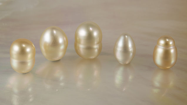
GIA’s Bangkok laboratory recently received five pearls ranging from cream to orangy yellow, weighing between 2.33 and 5.54 ct and measuring approximately 0.96 × 0.64 mm to 1.03 × 0.83 mm (figure 1). The pearls appeared similar to South Sea cultured pearls produced from Pinctada maxima mollusks. X-ray fluorescence imaging was used to confirm their saltwater origin; all samples were inert when exposed to X-rays, resulting from the absence of, or a very low level of, manganese, characteristic of saltwater pearls. The four orangy yellow pearls exhibited typical ultraviolet/visible/near-infrared reflectance spectra of natural color yellow South Sea pearls (S. Jin et al., “Application of UV-Vis-NIR spectroscopy to gemology,” Winter 2024 G&G, pp. 456–473).
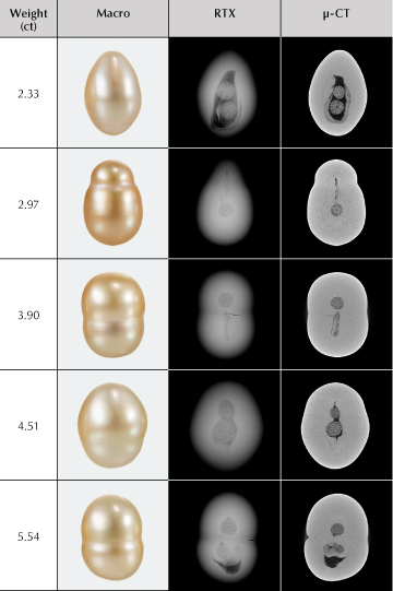
Real-time X-ray microradiography (RTX) analysis revealed that each sample contained one or two intriguing spherical radial objects in the center, positioned along the length of the pearl (figure 2). The objects were associated with varying forms of elongated void or linear features known to be diagnostic growth structures commonly found in South Sea non-bead cultured (NBC) pearls (A. Homkrajae et al., “Internal structures of known Pinctada maxima pearls: Cultured pearls from operated marine mollusks,” Fall 2021 G&G, pp. 186–205). The pearls were further analyzed using X-ray computed microtomography (μ-CT) to enhance image resolution, which provided a more comprehensive understanding of the internal structures.
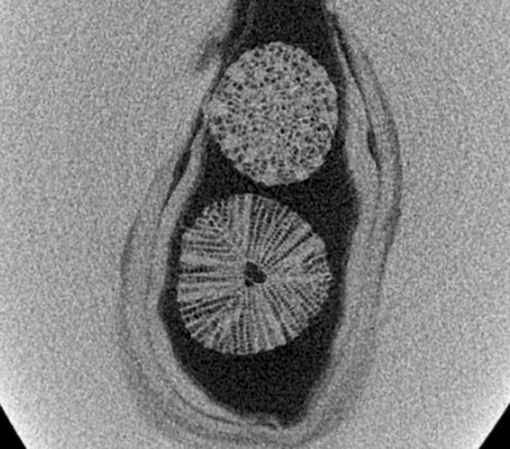
The spherical objects, along with the typical void and linear structures, were clearly visible in the μ-CT analysis. The 3D images revealed that each sphere possessed a distinctive porous and radial structure extending outward from the central core (figure 3; video coming soon). Their morphology closely resembled foraminifera spheres found in a natural pearl previously examined at GIA’s Bangkok laboratory (Winter 2015 Lab Notes, pp. 434–436). RTX analysis was used to obtain measurements of all the spherical objects, ranging from 1.23 to 2.28 mm in diameter, which are consistent with the size of previously reported microorganisms. However, the previously studied natural pearl exhibited dark organic- and conchiolin-rich growth layers around the rounded microorganism’s central core and growth arcs within the surrounding nacre, a structural characteristic comparable with the formation of natural pearls. Conversely, all five studied pearls displayed a tight structure or minimal growth arcs in the surrounding nacre, consistent with NBC pearls and the so-called “atypical bead cultured pearls” previously studied (“Atypical ‘beading’ in the production of cultured pearls from Australian Pinctada maxima,” GIA Research News, February 13, 2017).
Although previously reported microscopic marine organisms found within pearls were related to natural formations, the internal void and linear features of these studied samples, as well as their close resemblance to South Sea cultured pearls, indicated that these were likely of cultured origin. Moreover, it is very unusual to encounter similar-looking pearls with identical internal structures of microorganisms rarely found inside pearls. These findings indicated that microorganisms were intentionally inserted into the host mollusk as bead nuclei for culturing. The use of these organisms may be intended to imitate natural pearl formation; thus, gemologists must take caution when interpreting organic materials as evidence of natural origin, and comprehensive analysis is needed for identification. This is the first reported discovery of microscopic marine organisms in South Sea cultured pearls at GIA’s laboratories.
Kwanreun Lawanwong is a senior analytics technician at GIA in Bangkok.
GIA’s Bangkok laboratory recently received five pearls ranging from cream to orangy yellow, weighing between 2.33 and 5.54 ct and measuring approximately 0.96 × 0.64 mm to 1.03 × 0.83 mm (figure 1). The pearls appeared similar to South Sea cultured pearls produced from Pinctada maxima mollusks. X-ray fluorescence imaging was used to confirm their saltwater origin; all samples were inert when exposed to X-rays, resulting from the absence of, or a very low level of, manganese, characteristic of saltwater pearls. The four orangy yellow pearls exhibited typical ultraviolet/visible/near-infrared reflectance spectra of natural color yellow South Sea pearls (S. Jin et al., “Application of UV-Vis-NIR spectroscopy to gemology,” Winter 2024 G&G, pp. 456–473).
Real-time X-ray microradiography (RTX) analysis revealed that each sample contained one or two intriguing spherical radial objects in the center, positioned along the length of the pearl (figure 2). The objects were associated with varying forms of elongated void or linear features known to be diagnostic growth structures commonly found in South Sea non-bead cultured (NBC) pearls (A. Homkrajae et al., “Internal structures of known Pinctada maxima pearls: Cultured pearls from operated marine mollusks,” Fall 2021 G&G, pp. 186–205). The pearls were further analyzed using X-ray computed microtomography (μ-CT) to enhance image resolution, which provided a more comprehensive understanding of the internal structures.
The spherical objects, along with the typical void and linear structures, were clearly visible in the μ-CT analysis. The 3D images revealed that each sphere possessed a distinctive porous and radial structure extending outward from the central core (figure 3; video coming soon). Their morphology closely resembled foraminifera spheres found in a natural pearl previously examined at GIA’s Bangkok laboratory (Winter 2015 Lab Notes, pp. 434–436). RTX analysis was used to obtain measurements of all the spherical objects, ranging from 1.23 to 2.28 mm in diameter, which are consistent with the size of previously reported microorganisms. However, the previously studied natural pearl exhibited dark organic- and conchiolin-rich growth layers around the rounded microorganism’s central core and growth arcs within the surrounding nacre, a structural characteristic comparable with the formation of natural pearls. Conversely, all five studied pearls displayed a tight structure or minimal growth arcs in the surrounding nacre, consistent with NBC pearls and the so-called “atypical bead cultured pearls” previously studied (“Atypical ‘beading’ in the production of cultured pearls from Australian Pinctada maxima,” GIA Research News, February 13, 2017).
Although previously reported microscopic marine organisms found within pearls were related to natural formations, the internal void and linear features of these studied samples, as well as their close resemblance to South Sea cultured pearls, indicated that these were likely of cultured origin. Moreover, it is very unusual to encounter similar-looking pearls with identical internal structures of microorganisms rarely found inside pearls. These findings indicated that microorganisms were intentionally inserted into the host mollusk as bead nuclei for culturing. The use of these organisms may be intended to imitate natural pearl formation; thus, gemologists must take caution when interpreting organic materials as evidence of natural origin, and comprehensive analysis is needed for identification. This is the first reported discovery of microscopic marine organisms in South Sea cultured pearls at GIA’s laboratories.
Kwanreun Lawanwong is a senior analytics technician at GIA in Bangkok.