
A remarkable Brazilian pegmatite plate hangs on the ground-floor wall of the Centre for Petrographic and Geochemical Research (CRPG) in Vandœuvre, France. This rock, called “Patagonia granite” by the trade in reference to the granitic mountain peaks in southern Argentina, came from the Galiléia region, near Governador Valadares, in the state of Minas Gerais, Brazil. Famous for its gem-bearing pegmatites, the region, located north of Rio de Janeiro, is marked by the presence of granitoids belonging to the Neoproterozoic Galiléia magmatic suite formed by tonalites, granodiorites, granites, and pegmatites. Dating of these rocks by the uranium-lead method on zircon has given ages between 594 and 570 million years, characteristic of the Brasiliano or Pan-African orogeny.
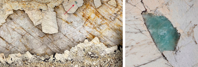
The polished pegmatite plate, measuring 3.28 m × 1.80 m × 2.5 cm, exhibits three distinct mineral assemblages from the border to the core (figure 1, left): (1) the edges composed of quartz, feldspar, and large brownish laths of mica (black in the photo) ending at the top with a close association of wedge-shaped quartz and potassium-feldspar (graphic texture); (2) large and zoned euhedral perthitic microcline feldspar crystals hosting disseminated centimetric schorlite and an unidentified 7.1 × 2.9 cm bluish green translucent mineral (figure 1, right); and (3) the whole core filled by translucent and healed grayish quartz.
The unidentified bluish green mineral was analyzed using visible/near-infrared (Vis-NIR) reflectance spectroscopy with an analytical point-spectrometer. The technique allows for the acquisition of a reflectance spectrum of a 1 cm diameter surface area. Spectra collected in the 500–2500 nm range are sometimes used to analyze the mineralogical composition of rocks based on the position and shape of the absorption band.
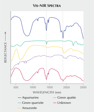
The spectrum of the bluish green mineral was compared with that obtained from sample standards in the CRPG database (amazonite, green apatite, aquamarine, and green quartzite; figure 2). The positions of the two absorption bands at 1400 nm and 1900 nm are linked to the vibration of water molecules in the structure, while the wide absorption band centered at 800 nm is due to an electronic transition of iron. Therefore, the unknown mineral was positively identified as a member of the beryl family.
Chemical analysis on the bluish green beryl by a handheld X-ray fluorescence (XRF) spectrometer showed the following analytical ranges for trace elements, in ppmw: iron = 3000 (±100), titanium = 447–474 (±27), zinc = 305–350 (±13), and cesium = 157–180 (±6). Chromium and vanadium were not detected, ruling out emerald and identifying the mineral as aquamarine, with its color attributed to the combination of iron and titanium. The presence of cesium confirmed the magmatic source of the aquamarine.
The determination of aquamarine on the large plate of Brazilian pegmatite combining handheld Vis-NIR reflectance and XRF spectrometers underscores the importance of the use of nondestructive methods on polished petrological sections in galleries of mineralogical museums. These qualitative point chemical analyses offer insight into the knowledge of mineral associations across diverse geological environments and enhance understanding of the genesis of gems.
Gaston Giuliani, Marie Barthez, Jean-Marc Montel, and Pierre Barbey are affiliated with the Université de Lorraine, CRPG/CNRS-UL, in Vandœuvre, France.
A remarkable Brazilian pegmatite plate hangs on the ground-floor wall of the Centre for Petrographic and Geochemical Research (CRPG) in Vandœuvre, France. This rock, called “Patagonia granite” by the trade in reference to the granitic mountain peaks in southern Argentina, came from the Galiléia region, near Governador Valadares, in the state of Minas Gerais, Brazil. Famous for its gem-bearing pegmatites, the region, located north of Rio de Janeiro, is marked by the presence of granitoids belonging to the Neoproterozoic Galiléia magmatic suite formed by tonalites, granodiorites, granites, and pegmatites. Dating of these rocks by the uranium-lead method on zircon has given ages between 594 and 570 million years, characteristic of the Brasiliano or Pan-African orogeny.
The polished pegmatite plate, measuring 3.28 m × 1.80 m × 2.5 cm, exhibits three distinct mineral assemblages from the border to the core (figure 1, left): (1) the edges composed of quartz, feldspar, and large brownish laths of mica (black in the photo) ending at the top with a close association of wedge-shaped quartz and potassium-feldspar (graphic texture); (2) large and zoned euhedral perthitic microcline feldspar crystals hosting disseminated centimetric schorlite and an unidentified 7.1 × 2.9 cm bluish green translucent mineral (figure 1, right); and (3) the whole core filled by translucent and healed grayish quartz.
The unidentified bluish green mineral was analyzed using visible/near-infrared (Vis-NIR) reflectance spectroscopy with an analytical point-spectrometer. The technique allows for the acquisition of a reflectance spectrum of a 1 cm diameter surface area. Spectra collected in the 500–2500 nm range are sometimes used to analyze the mineralogical composition of rocks based on the position and shape of the absorption band.
The spectrum of the bluish green mineral was compared with that obtained from sample standards in the CRPG database (amazonite, green apatite, aquamarine, and green quartzite; figure 2). The positions of the two absorption bands at 1400 nm and 1900 nm are linked to the vibration of water molecules in the structure, while the wide absorption band centered at 800 nm is due to an electronic transition of iron. Therefore, the unknown mineral was positively identified as a member of the beryl family.
Chemical analysis on the bluish green beryl by a handheld X-ray fluorescence (XRF) spectrometer showed the following analytical ranges for trace elements, in ppmw: iron = 3000 (±100), titanium = 447–474 (±27), zinc = 305–350 (±13), and cesium = 157–180 (±6). Chromium and vanadium were not detected, ruling out emerald and identifying the mineral as aquamarine, with its color attributed to the combination of iron and titanium. The presence of cesium confirmed the magmatic source of the aquamarine.
The determination of aquamarine on the large plate of Brazilian pegmatite combining handheld Vis-NIR reflectance and XRF spectrometers underscores the importance of the use of nondestructive methods on polished petrological sections in galleries of mineralogical museums. These qualitative point chemical analyses offer insight into the knowledge of mineral associations across diverse geological environments and enhance understanding of the genesis of gems.
Gaston Giuliani, Marie Barthez, Jean-Marc Montel, and Pierre Barbey are affiliated with the Université de Lorraine, CRPG/CNRS-UL, in Vandœuvre, France.