Natural Shell Blisters and Blister Pearls: What’s the Difference?
Introduction
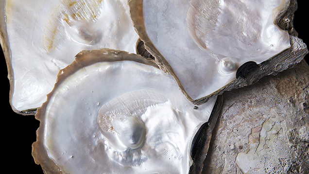
The majority of saltwater and freshwater pearls bought and sold in the global jewelry markets are “whole pearls,” sometimes referred to as “cyst pearls” (CIBJO, 2017). These pearls form within the soft tissue of a mollusk, either naturally or with human intervention as complete pearls, hence the term “whole.” Such pearls are usually more desirable and command higher prices than those classified as “blister pearls.” While not technically a pearl, there is a third type of formation known as a “shell blister” or “blister” (Strack, 2006) that needs to be considered when addressing this subject. This article addresses the differences between these three materials, with a focus on natural shell blisters and natural blister pearls (figure 1). In order to fully understand these two organic formations, it is helpful to first discuss a natural cyst (whole) pearl in more detail.
A natural cyst (whole) pearl starts forming within a mollusk’s soft tissue and remains in the pearl sac until the time that it is removed (figure 2). While some gemologists believe that this definition is the only one that should be applied to natural cyst pearls, others consider any natural pearl that breaks through the mantle and starts to transition into a natural blister pearl, even when not covered with enough shell layers to make the pearl and shell one entity a natural cyst pearl. The latter type is sometimes found on a shell’s inner surface and may be freed with sufficient pressure from the fingers. No form of cutting or grinding is required in such cases.
 pearl formation.")
A natural blister pearl forms naturally as a whole pearl within a pearl sac in the mantle (see natural cyst pearl). The natural pearl breaks through the sac and eventually finds its way between the shell and mantle. Nacreous or non-nacreous layers of calcium carbonate are secreted over the pearl and attach it to the shell (CIBJO, 2017). The completely merged entity is now by definition a natural blister pearl (figure 3). As the mollusk grows older, the thickness of the enveloping layers increases. Unlike whole pearls, these are removed from their shells through cutting and grinding in order to be suitable for use in jewelry as loose pearls. Some smaller shells with blister pearls are incorporated in pieces of jewelry with unique designs.

A natural shell blister is an internal protuberance that forms on a shell’s inner surface. It is usually caused by a foreign object accidentally finding its way into the space between the mantle and shell surface. The mollusk attempts to alleviate this irritation with the secretion of nacreous layers (figure 4). The foreign object may be a shell-boring parasitic worm or sponge (CIBJO, 2017), or perhaps some other organic or inorganic material that found its way into the shell while the mollusk was feeding.

Materials and Methods
A total of 33 samples were selected for this study, including 7 natural cyst pearls (all were lightly attached to their host shell at one stage), 18 natural blister pearls, and 8 natural shell blisters. They formed on Pinctada maxima mollusks recovered by GIA gemologists during field trips in Broome, Australia, hosted by Paspaley Pearling Company. All the specimens were obtained off 80 Mile Beach during operations to fish for wild Pinctada maxima shell as a part of the Australian shelling quota system (WAMSC, 2015). The measurements of the cyst pearls ranged from 4.52 × 4.40 × 3.02 mm to 16.23 × 15.65 × 13.21 mm. The natural blister pearl specimens ranged from approximately 5.92 × 3.85 mm to 18.32 × 14.08 mm, and the natural shell blister samples ranged from approximately 7.80 × 5.09 mm to 32.90 × 23.85 mm. The dorsoventral (DVM) measurements of the shells ranged from approximately 151.20 × 134.46 mm to 184.75 × 172.68 mm. The photos, photomicrographs, and microradiographs of the all samples are shown in tables 1–3.
Routine pearl testing methods were used to examine the specimens, with microradiography the main technique used to examine their internal features. A Pacific X-ray Imaging (PXI) GenX-90P real time X-ray (RTX) machine with 4 micron microfocus, 90 kV voltage, and 0.18 mA current X-ray source fitted with a PerkinElmer flat panel detector was used to reveal the internal structures.
Photomicrographs showing detailed surface features of the natural cyst (whole) pearls where they made contact with the shells, as well as natural blister pearls and natural shell blisters attached to their shells, were taken using a Nikon SMZ18 stereo-microscope with an SHR Plan Apo 1× objective lens and NIS-Elements imaging software.
Results
One of the most important ways to examine a pearl is through the visual examination of its external features, first with the unaided-eye, followed by a loupe, and then a microscope for more detail. The other is the examination of the internal structure using microradiography (RTX) and, when necessary and if possible, X-ray computed microtomography (µ-CT). When it comes to identifying natural blister pearls and natural shell blisters, these techniques are especially important as it is not always simple to make a clear separation. However, the use of µ-CT analysis in the examination of any pearl still attached to a large shell sample is not practical, and thus the results usually depend on RTX analysis.
Natural Cyst Pearls (Contact with Their Shells)
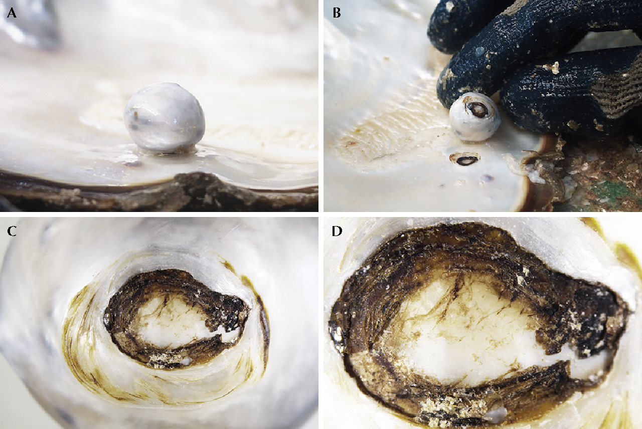
If a natural blister pearl forms in the usual manner (figure 3), visually identifying it as a blister pearl can be difficult since the original pearl is covered by the overlying layers. However, if the pearl is harvested before the mollusk deposits additional layers, it may still be classified as a whole pearl even if contact has been made with the shell (figure 5A). In such examples, the original shape of the pearl with a limited contact area with the shell is usually apparent. Natural cyst pearl A-1 was easily removed from its host shell by applying gentle pressure with the fingers of one hand (figure 5, A and B). A tell-tale contact point of brown organic rich rings was visible after removal on both the pearl and shell (figure 5, C and D). A natural blister pearl or natural shell blister would in contrast have to be cut from its host shell. Such examples usually show the resulting work and/or polishing on the base, at the very least, as evidence to support the action. The internal structure of the pearl in figures 5 and 6A revealed characteristic growth features expected for natural pearls in the way of a small dark organic-rich core in the center with clear associated concentric growth structures (figure 6, B and C).
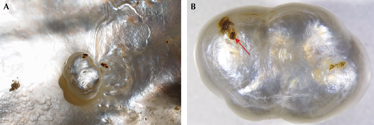
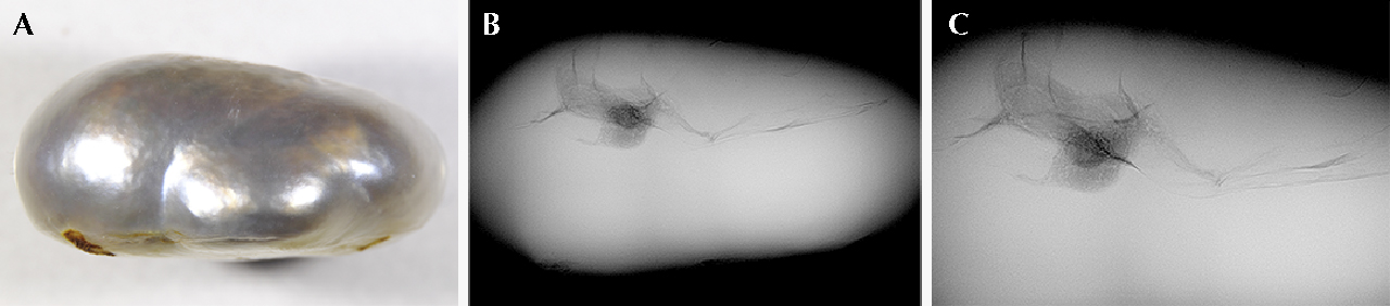
However, it is possible to find a whole pearl that was in contact with its host but lacks any convincing signs of contact with the shell (e.g., pearl A-2 which only shows minimal organic-rich features on the base; figure 7, A and B). This type of pearl would not be considered a natural blister pearl if submitted “blind” (without any details about its history) for testing. Sometimes the internal structures encountered in natural cyst pearls may be unusual. Pearl A-2 (figure 8A) shows a suspect linear feature with an associated void/organic-rich structure (figure 8, B and C) that may be considered more representative of non-bead cultured pearls (Krzemnicki et al., 2010; Sturman et al., 2016; Nilpetploy et al., 2018a). However, the pearl in question is undoubtedly natural despite its suspect internal structure.
In the majority of cases, it is not difficult to comprehend that the RTX structures of cyst pearls that do make contact with the shell look very much like those that form in the body of the animals. In some examples, a very small to moderately sized contact point may be the only evidence of contact visible on the pearl’s surface, but it is not always possible to be certain whether the area is shell related or the result of another pearl (twin or multiple) once being attached.

Natural Blister Pearls
Natural blister pearls may potentially be found on the shells of any mollusk, but the majority of saltwater natural blister pearls and natural shell blisters form within the mollusks of the Pteriidae family. This is shown by the number of such pearls and shell blisters that pass through GIA’s laboratories on an annual basis. The largest of these semi-baroque to baroque features may measure over two centimeters in length and tend to form specifically within Pinctada maxima mollusks.
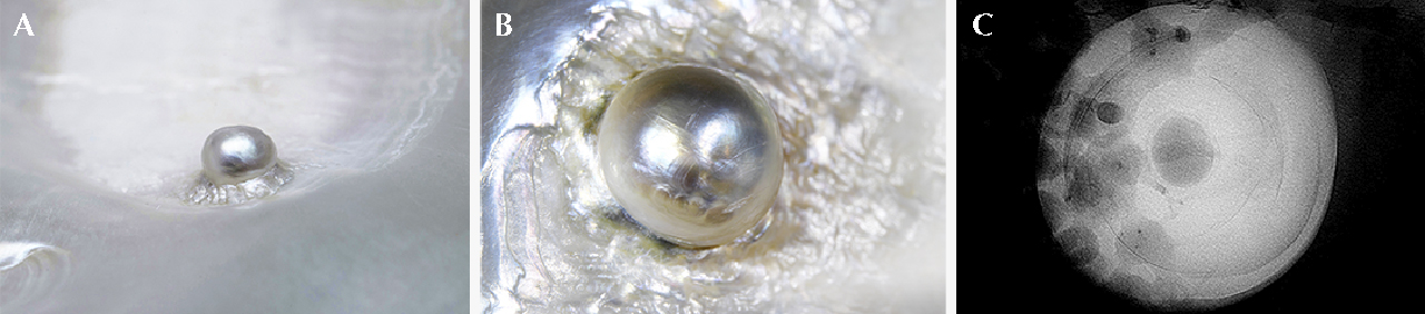
Typically, the structures of natural blister pearls, as revealed by RTX analysis, differ from those associated with natural cyst pearls overgrown with additional layers of material. Examples of natural blister pearl structures from a number of different mollusks have already been recorded in the literature (Nilpetploy et al., 2018b; Ho, 2018). Most such samples in this study exhibited straightforward natural internal growth structures. Four examples with a variety of natural structures are shown in greater detail in figures 9–12. A blister pearl (B-7) shows a dark organic-rich nucleus surrounded by concentric growth arcs via RTX analysis (figure 9C). Also note the rippled nacreous layers around the pearl (figure 9, A and B) that secure it to the host shell. Sample B-1 (figure 10) is a pearl with a near-drop shaped outline that is attached to a shell. The RTX results revealed concentric growth arcs that are difficult to resolve since they overlap with dark parasitic channel structures beneath the pearl within the shell itself (figure 10, B and C). Sample B-11 (figure 11) clearly illustrates where multiple pearls have joined together to form an aggregate pearl on the shell’s surface. The boundaries between each individual pearl segment may be seen as dark folds cutting across the pearl (figure 11B, yellow arrows). Such aggregate examples often reveal natural internal structures within each segment (figures 11, C-F). Clear concentric growth arcs are shown in the segment analyzed in figure 11C. The final sample (B-2) shows organic-rich or void-like features more in keeping with natural shell blisters (figure 12). However, the general external form (height/depth, shape, outline) are more in keeping with those expected for natural blister pearls based on the authors’ experience.


Natural Shell Blister
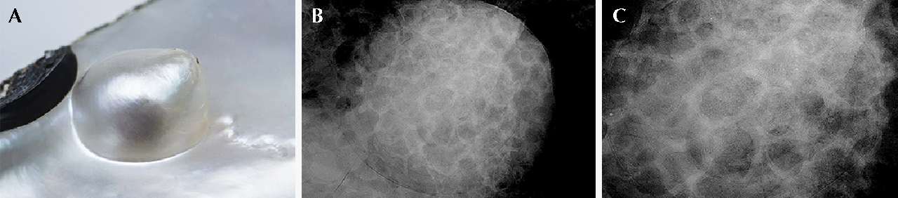
In many examples, natural shell blisters show a form similar to natural blister pearls since they appear to be nacreous concretions protruding from the inner wall of a shell. However, the majority do not show a pronounced boundary around the protrusion and are often more “shallow” than natural blister pearls. Additionally, natural shell blisters appear to “blend in” more with their host and show as bumps in which the overlying CaCO3 “overflows,” such as illustrated by sample C-4 (figure 13A). Many natural shell blisters are large and more importantly tend to show organic-rich features, often with partial or significant hollow structures (Manustrong, 2016). The voids might well be due to the decomposition of organic matter that instigated the formation of the blister. When drilled, such examples may produce a disagreeable smell as the fermented gases escape (Sturman, 2009a).
Three natural shell blister samples attached to their host shells are shown in figures 13–15. Only sample C-1 (figure 15A) shows a partial blister pearl form (boundary and attachment area on one side stand proud of the shell). However, the overall appearance indicates it is more likely a natural shell blister. The internal structures of all three reveal partially hollow structures, the darker veins or patches resulting from underlying parasite channels within the shell rather than the blister structure itself (figures 13B, 14B, and 15B). Some growth arcs are visible in areas around the organic-rich/void features indicating the blisters formed naturally.
The examination of natural blister pearls and natural shell blisters attached to their shells is limited by the number of directions in which they may be examined within the RTX unit. This is a restriction that does not come into play with loose pearls or blisters that have been removed from their shells by sawing and working. Due to the size of the whole shell and position of the target, often somewhere near the center, the usual scenario is that only one direction is possible, that being the direction shown in figures 9–13. In some rare cases, such as the shell blisters in figure 14 and 15, it is possible to inspect the target from its side. The results clearly show the thicker curved solid outline of the blister and darker more radio-transparent organic rich/hollow structure within (figures 14C and 15C). The black “vein” visible in figures 15B and 15C (indicated by red arrows) appears to be the same parasite tube showing in different orientations. Occasionally, RTX analysis may reveal the existence of the organism that initiated the formation of a natural shell blister. Fish and small crabs have all been recorded in previous literature (Edwards, 1913; Chan and Yasawa, 2018), however, some species of shrimp are also known to co-exist with the former two creatures and could potentially be the instigators of natural shell blister formations (McLaurin, 2010). In some cases, opinions on the correct nomenclature to describe the entombed type of organisms arises, and these differences make their way into the literature as illustrated by the two cases described in Hainschwang (2009) and Chan and Yasawa (2018).


Conclusion
All the nacreous natural cyst pearl, natural blister pearl, and natural shell blister samples in this study were of known natural origin since the shells were obtained directly from the sea off 80 Mile Beach, North Western Australia, far away from any cultured pearl farms. The majority of the natural cyst pearls and natural blister pearls showed natural pearl structures consisting of concentric growth rings or arcs toward their centers with a generally solid aspect to their microradiographic appearance (white, more radio-translucent form), whereas the majority of natural shell blisters revealed varying degrees of organic-rich or void associated structures illustrating the less-solid forms of blisters. Laboratory experience has shown that these results tend to be repeatable in the majority of natural blister pearls and natural shell blisters, in that the former almost always appear solid and therefore whiter when examined in GIA’s RTX units, while the latter show void-like or less-solid areas. Sometimes an organic-rich feature may show in alignment with the position of a natural blister pearl and appear as though it is within the blister pearl itself. However, if we were able to examine the blister pearl at right angles to the first orientation it would soon become clear that the organic-rich area is positioned within the shell component beneath the natural blister pearl and is not associated in any way with the blister pearl itself. Unfortunately, the repositioning and examination of natural blister pearls attached to their hosts is not possible owing to the potential damage of the shell’s rim, especially if nearer the lip area. The quality of the resulting RTX images also tends to be impacted by the position of the target and the obstacles (shell ridges/features, other blisters, or blister pearls, etc.) that may be in the way.
Another useful observation that may help differentiate between natural blister pearls and natural shell blisters is that blister pearls almost always appear to be like whole pearls that have attached themselves to the shell. The height of the domes (thicknesses) are greater than those generally seen in natural shell blisters which seem to merge more with their host shells forming shallower “bumps” on the surface.
While the identification of most natural blister pearls and natural shell blisters is relatively straightforward, this is certainly not always the case and sometimes even experienced gemologists disagree (Zwaan et al., 2014; Krzemnicki and Braun, 2017; Dommisse, 2018). As the auction example referenced clearly illustrates, the problem is often made even harder when work has been carried out to remove the feature from its host. This changes the surface condition and shape of the object being analyzed. The work may be fairly minor and just involve cutting the base area away to separate it from the shell. Or the work may be more significant in that it alters not only the base but also the sides of the piece. If heavily worked, even the entire surface may be altered. In addition to such work, the various steps of “working” (cutting, grinding, and smoothing) the item is often followed by varying degrees of polishing to improve the dull/matte appearance. Significant polishing, referred to as “heavily polished” on GIA pearl reports, is also a factor that may alter the surface to quite a degree and is used to mask the worked areas of the item under examination.
While this article has only addressed the topic in relation to nacreous pearls, it should be noted that all mollusks have the potential to produce natural blister pearls and natural shell blisters (Sturman, 2009b; Zhou et al., 2015; Nilpetploy et al., 2018b; Ho, 2018). This is clearly evident in the numerous reports in the media about “natural blister pearls” from the Tridacna gigas (giant clam) and other species from the Tridacninae subfamily (Beavers, 2018). Many of these eventually turn out to be shell that has been heavily worked, and in most cases polished or heavily polished, to appear as natural blister pearls (Krzemnicki and Cartier, 2017). Some may also be considered natural shell blisters rather than natural blister pearls, but it is GIA’s experience that the vast majority are pieces of fashioned shell.


